

MRI might be considered a mature modality, but creative and thoughtful researchers and clinicians are constantly exploring new avenues for the decades-old imaging technology.
Multiparametric MRI, hybrid multidimensional MRI, newly developed sequences for metal artifact reduction, and texture analysis are just a sampling of the advances designed to benefit patients for a wide variety of clinical indications.
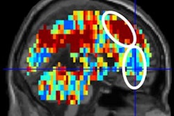
In addition, functional MRI continues to play a prominent role in unlocking secrets of the brain -- what causes us to think and act the way we do, how we recover from concussions, and why dementia and Alzheimer's disease have become such devastating conditions.
At the same time, MRI has its share of controversy. Many practitioners rightly view the modality as an expensive imaging option. Here again is where resourcefulness leads to imaginative approaches. This year's RSNA 2017 agenda offers strategies to shorten scan times to reduce costs and yield diagnostic results, while appropriate use criteria provide a direct and informed path to improved outcomes.
The safety of gadolinium-based contrast agents (GBCAs) again will take center stage at RSNA 2017. The discussion continues regarding how gadolinium retention occurs in many areas of the body and to what extent that accumulation may or may not affect patients' long-term health.
Could ferumoxytol-enhanced MRI be the solution? How safe are MRI scans in patients with implantable cardiac electronic devices? The answers to those questions and more are available in the summaries of MRI-related scientific presentations and open forums below.
To view the RSNA's complete listing of abstracts for the 2017 scientific and educational program, click here.

![Overview of the study design. (A) The fully automated deep learning framework was developed to estimate body composition (BC) (defined as subcutaneous adipose tissue [SAT] in liters; visceral adipose tissue [VAT] in liters; skeletal muscle [SM] in liters; SM fat fraction [SMFF] as a percentage; and intramuscular adipose tissue [IMAT] in deciliters) from MRI. The fully automated framework comprised one model (model 1) to quantify different BC measures (SAT, VAT, SM, SMFF, and IMAT) as three-dimensional (3D) measures from whole-body MRI scans. The second model (model 2) was trained to identify standardized anatomic landmarks along the craniocaudal body axis (z coordinate field), which allowed for subdividing the whole-body measures into different subregions typically examined on clinical routine MRI scans (chest, abdomen, and pelvis). (B) BC was quantified from whole-body MRI in over 66,000 individuals from two large population-based cohort studies, the UK Biobank (UKB) (36,317 individuals) and the German National Cohort (NAKO) (30,291 individuals). Bar graphs show age distribution by sex and cohort. BMI = body mass index. (C) After the performance assessment of the fully automated framework, the change in BC measures, distributions, and profiles across age decades were investigated. Age-, sex-, and height-adjusted body composition reference curves were calculated and made publicly available in a web-based z-score calculator (https://circ-ml.github.io).](https://img.auntminnie.com/mindful/smg/workspaces/default/uploads/2026/05/body-comp.XgAjTfPj1W.jpg?auto=format%2Ccompress&fit=crop&h=100&q=70&w=100)


![Overview of the study design. (A) The fully automated deep learning framework was developed to estimate body composition (BC) (defined as subcutaneous adipose tissue [SAT] in liters; visceral adipose tissue [VAT] in liters; skeletal muscle [SM] in liters; SM fat fraction [SMFF] as a percentage; and intramuscular adipose tissue [IMAT] in deciliters) from MRI. The fully automated framework comprised one model (model 1) to quantify different BC measures (SAT, VAT, SM, SMFF, and IMAT) as three-dimensional (3D) measures from whole-body MRI scans. The second model (model 2) was trained to identify standardized anatomic landmarks along the craniocaudal body axis (z coordinate field), which allowed for subdividing the whole-body measures into different subregions typically examined on clinical routine MRI scans (chest, abdomen, and pelvis). (B) BC was quantified from whole-body MRI in over 66,000 individuals from two large population-based cohort studies, the UK Biobank (UKB) (36,317 individuals) and the German National Cohort (NAKO) (30,291 individuals). Bar graphs show age distribution by sex and cohort. BMI = body mass index. (C) After the performance assessment of the fully automated framework, the change in BC measures, distributions, and profiles across age decades were investigated. Age-, sex-, and height-adjusted body composition reference curves were calculated and made publicly available in a web-based z-score calculator (https://circ-ml.github.io).](https://img.auntminnie.com/mindful/smg/workspaces/default/uploads/2026/05/body-comp.XgAjTfPj1W.jpg?auto=format%2Ccompress&dpr=2&fit=crop&h=167&q=70&w=250)


