Dear Ultrasound Insider,
Three-dimensional ultrasound has demonstrated clinical utility across a range of applications, and the technique is also useful in thigh studies, according to a case study provided to AuntMinnie.com by Dearbhla O'Dwyer and Dr. Stefano Ciatti of the Studio Ecograficao Dott. Stefano Ciatti in Prato, Italy.
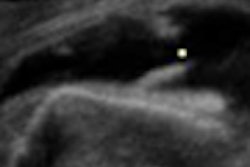
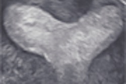
In a case involving a complete tear of the rectus femoris muscle in an adolescent soccer player, ultrasound -- including multiplanar imaging and 3D reconstruction -- provided valuable information about the condition of the thigh muscle rupture. Gaining access to the coronal plane allows for better appreciation of tears, according to O'Dwyer and Ciatti.
As an Ultrasound Insider subscriber, you have access to this Insider Exclusive before it is published for the rest of our AuntMinnie.com members. To learn more about ultrasound in the thigh, click here.
While you're visiting your Ultrasound Digital Community, be sure to check out our other current articles, including a story on recent research evaluating thyroid ultrasound. We're also featuring continuing coverage from last month's Leading Edge in Diagnostic Ultrasound conference, including stories on how oral contrast agents show potential for gastrointestinal applications and how ultrasound contrast boosts prostate biopsy yield.
In addition, read about how screening ultrasound boosts the breast cancer detection rate in high-risk women. You can also find out why ultrasound should be the first imaging study for suspected appendicitis by clicking here.
Have an idea for a topic you'd like to see covered? As always, please feel free to drop me a line.


