Most women who require a diagnostic workup based on mammographic screening results do not have cancer. But there are instances when a diagnostic exam will turn up a cancer diagnosis in another area of the breast that was not a clinical concern initially. In addition, up to 2.6% of patients with a newly diagnosed breast cancer also have a second lesion, either in the same or in the contralateral breast.
Radiologists from two branches of Kaiser Permanente in California set out to determine if computer-aided detection (CAD) could enhance diagnostic mammograms, potentially picking up these secondary cancers. They shared their results in the November issue of the American Journal of Roentgenology.
"We defined diagnostic mammograms as standard four-view mammograms obtained in patients who presented with clinical signs or symptoms that aroused suspicion of breast cancer," wrote lead author Dr. Sherry Butler from Kaiser Permanente in San Francisco. Two of the co-investigators are from the Kaiser Permanente in Redwood City (AJR, November 2004, Vol. 183:5, pp.1511-1515).
For the retrospective review, the group collected data from the two facilities, as well as from a Kaiser Permanente in Santa Clara, CA. A total of 197 patients with 212 biopsy-proven cancers made up the final patient population. Diagnostic mammograms were reviewed with the ImageChecker M1000, version 2.2 (R2 Technology, Sunnyvale, CA). Two additional study co-authors are affiliated with the CAD company.
 |
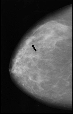
| Fifty-eight-year-old woman who presented with "pulling" on the inner left breast, diagnosed with bilateral invasive ductal carcinoma after biopsy. Above is the craniocaudal view mammogram. Cancer in left breast at point of clinical finding is apparent. A second cancer (arrow) was detected in the right breast. Butler SA, Gabbay RJ, Kass DA, Siedler DE, O'Shaughnessy KF, Castellino RA, "Computer-Aided Detection in Diagnostic Mammography: Detection of Clinically Unsuspected Cancers," (AJR 2004; 183: 1511-1515). |
A total of 761 images were reviewed. The most common clinical finding that prompted a diagnostic mammogram was a palpable mass (90%). A single biopsy-proven cancer was identified in 92.4% of the women with an additional cancer at a second site found in 7.6%. These clinically unsuspected cancers found at an unrelated site -- 30 in total -- appeared on mammography as either mass, architectural distortion, or microcalcification clusters. The 30 cancers allowed a measurement of the potential benefit of CAD, the authors wrote.
 |
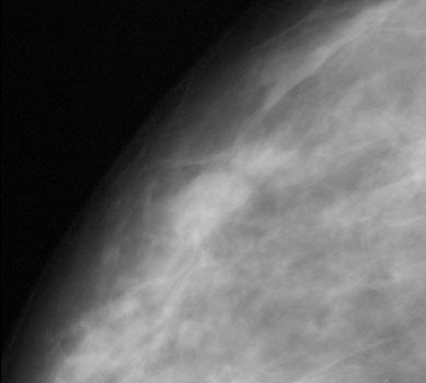
| Same patient. A digital magnification of the image above. Cancers in both left and right breasts were marked by CAD system. Butler SA, Gabbay RJ, Kass DA, Siedler DE, O'Shaughnessy KF, Castellino RA, "Computer-Aided Detection in Diagnostic Mammography: Detection of Clinically Unsuspected Cancers," (AJR 2004; 183: 1511-1515). |
In the end, the CAD system marked 87% of the biopsy-proven secondary cancers. More specifically, it caught 88% of the masses and 85% of the microcalcification clusters. The average number of false per four-view mammogram was 2.7, higher than the 2.0 reported for previous studies that tested CAD in a screening setting, the authors wrote. This is most likely explained by the fact that most known cancers are more complex, and under greater scrutiny by the CAD system, they wrote.
Fifteen percent of the diagnostic mammograms in this study had another cancer detected with CAD. "The use of CAD systems to help direct the attention of the radiologist to all suspicious areas in the image could be a potential benefit," the group concluded.
However, they acknowledged that they "have no proof that a radiologist prospectively using the CAD system would take action on the basis of one of the CAD marks." Prospective evaluations are required in this diagnostic setting, they wrote.
For more research on breast-related CAD, check out the following presentations at the 2004 Radiological Society of North America (RSNA) meeting in Chicago:
"The Potential Role of CAD in the Earlier Detection of Prior Cancers in Mammography," Poster 0306BR-p.
"Clinical Utilization of Computer-Aided Diagnosis in Mammography," Refresher Course RC625B.
"Usefulness of Similar Images for Distinction Between Benign and Malignant Lesions in Mammograms: Determination of Similarity between Pairs of Mammographic Lesions," Education Exhibit 0047PH-CE-e.
"Breast Computer-Aided Diagnosis: Limitations and Future Directions," Refresher Course RC625C.
By Shalmali Pal
AuntMinnie.com staff writer
October 27, 2004
Related Reading
Computer-aided diagnosis may reduce unnecessary breast biopsy, October 14, 2004
Computer-aided detection of screening mammograms curbs false negatives, July 23, 2004
R2 closes U.S. government deal, May 27, 2004
Copyright © 2004 AuntMinnie.com


