A radiologist’s perception when viewing a complex MR image may be akin to a Major League Baseball (MLB) batter reading the stitches on a fastball, according to researchers exploring exactly how diagnostic interpretations are made.
The baseball metaphor works because eye-tracking studies have shown that radiologists are able to discriminate between normal and abnormal stacks of 26 T2-weighted images from prostate MRI in as little as 48 milliseconds per section, said neuroscientist Robert Alexander, PhD, of the New York Institute of Technology in New York City, in an interview with AuntMinnie.com.
Similarly, expert batters discriminate between different types of pitches (eg, a curve ball vs. a fastball) in under 200 milliseconds, he noted.
“We know that if you do have a very high level of expertise at a task, you can get to a point where it’s almost astounding how quickly you can process things,” Alexander said.
Robert Alexander, MD, of SUNY Downstate Health Sciences University in Brooklyn, recently discussed with AuntMinnie.com what it means to be an expert reader in radiology, with the concept under investigation.
Alexander was co-lead author of a significant paper published in 2022 on mandating limits on workload, duty, and speed in radiology. He said complicating these investigations is that trainees have been shown to perform as well at times as expert radiologists.
In August last year, Alexander and colleagues conducted a psychophysical and eye-tracking study aimed at quantifying the gaze dynamics used by professional radiologists to detect abnormalities in medical images.
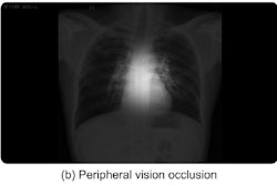
The study involved nine individuals with no medical imaging experience, 11 radiology residents, and six attending radiologists who were tasked with interpreting chest x-rays, each of which contained a potentially cancerous nodule. They found that some observers performed better than expected for their rank and years of experience.
“These results highlight that, at best, experience is an uncertain predictor of expertise level,” Alexander said.
Home runs or fly balls?
With this science in mind, Alexander and colleagues – notably Dr. Jeremy Wolfe, an ophthalmologist and radiologist at Harvard Medical School -- reprised the Perception Lab at ECR 2024 in Vienna, as well as presented ongoing research. The Perception Lab is a "pop-up" version of an academic research lab focused on medical image perception.
Nine labs from around the U.S. and Europe offered ECR attendees opportunities to participate in studies, with about 400 people tested, according to Wolfe.
“Sometimes out of these little experiments can come much bigger studies where you actually try to change practice in a clinical setting,” he said.
To that end, Wolf presented the group’s latest research in a session on perception related to why findings are sometimes missed on medical imaging. The researchers use eye-tracking technology to analyze the scan paths of radiologists as they view images to try to understand the mechanism underlying image perception.
According to the methodology, there are three types of errors:
- “Search” errors, in which the eye of the radiologist never lands on the target – a lesion on a mammogram, for instance
- “Recognition” errors, in which the eye lands near the target but leaves within 500 milliseconds
- “Decision” errors, in which the target is scrutinized, yet not reported
 A continuum of errors related to image perception. Image courtesy of Jeremy Wolfe, MD.
Together, these errors may be called the “Kundel continuum,” based on pioneering research on medical image perception by Harold Kundel, MD, a professor emeritus of radiology at the University of Pennsylvania.
When search errors and recognition errors intersect, they are called perception errors, Wolfe said.
“These are what we would call ‘look but fail to see’ errors. You really looked at it, but you somehow didn't succeed in processing it successfully,” he said.
One contributing factor involved in these errors may be incomplete sampling, he said. For instance, the eye-tracking data reveal that even if a radiologist is looking very near to the target, there is only a 50% chance that they will fixate on the target next, he said.
“Even when you are very nearby, you don't process everything successfully that's nearby and you may move off without ever looking at it,” he said.
Strategies for helping prevent these errors could involve checklists, double reading, and eventually sensible application of AI, Wolfe suggested.
Legal implications
Aside from the presentation, Wolfe noted that the research may have implications in medicolegal settings, where in the U.S., radiologists are often sued for missing findings on images. It can be very difficult in court to convince a juror that the miss wasn’t due to a misread when a lawyer can point to the finding and say, “Even I can see this,” he said.
“A lot of the very basic research that we do in my lab back in Boston is devoted to trying to figure out why it is that people, even when they're being diligent and they are expert, will still miss some things,” he said.
Ultimately, there are many unknowns, yet intuition may be at the heart of the mystery, Wolfe suggested.
“Everybody has good intuitions. The intuitions don’t necessarily agree with each other, and even if they did, they might not be right. So, we do these experiments because the question is interesting and because we can’t just trust our intuitions about what the right answer is,” he concluded.