Infectious Aortic Pseudoaneurysm:
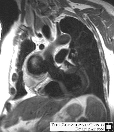
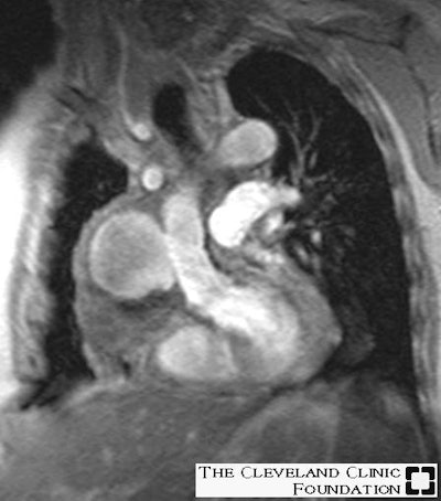
Below are images of a pseudoaneurysm at the distal anastamosis of an ascending aortic homograft that occurred secondary to infection. Both images were obtained in a double oblique coronal projection. The first image is a fast spin-echo T2-weighted sequence showing the large pseudoaneurysm that contains a mild amount of thrombus along the periphery, while the second image is a cine gradient-echo demonstrating the wide (3 cm) communication between the pseudoaneurysm and the ascending aorta.


