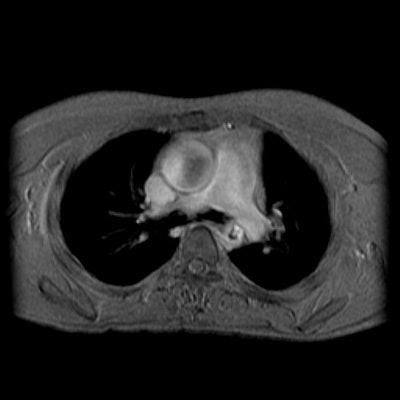
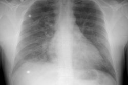
Aortic Coarctation:
The patient below was noted to have asymmetric pulses between the upper and lower extremities. The off-axis sagittal T1 weighted image demonstrates a focal narrowing of the descending thoracic aorta just beyond the ligamentum arteriosum. (Click on images to enlarge)
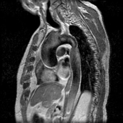
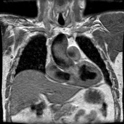
There is prominence to the ascending aorta seen on the coronal image. The patient was found to have a bicuspid aortic valve and this is likely related to post-stenotic dilatation of the ascending aorta.
Flow images reveal signal void in the descending aorta at the level of stenosis consistent with turbulent flow. Some signal void is also noted in the ascending aorta- likely related to the bicuspid valve.
