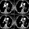
Re-expansion Pulmonary Edema:
Clinical:
Reexpansion pulmonary edema is a rare complication secondary to the rapid reexpansion of a chronically collapsed lung. In most cases the duration of collapse is greater than 3 days. The condition is usually associated with the use of high negative intrapleural pressures (greater than -20 cm of water) during reexpansion. Reexpansion pulmonary edema is likely multifactoral and related to prolonged local hypoxia, abrupt restoration of pulmonary blood flow, and sudden increase in marked negative intrapleural pressure [3]. The condition usually has a dramatic presentation- occurring immediately or within one hour in 64% of patients, and within 24 hours in the remainder. The clinical manifestations are varied ranging from asymptomatic to cardiorespiratory insufficiency. Nearly three-quarters of patients will have some form of symptoms. Symptoms typically begin 15 minutes to 2 hours after reexpansion and include dyspnea, tachypnea, cyanosis, cough, and unilateral rales on auscultation.Gradual reexpansion of a collapsed lung is appropriate if the duration of collapse exceeds 3 days. The risk of reexpansion pulmonary edema is minimized by the slow, staged removal of fluid not to exceed 1 liter over several hours and by the slow evacuation of a pneumothorax employing an underwater seal without suction. In patients with large effusions, only 1200 ml of fluid should be withdrawn during a single thoracentesis and patients should undergo careful observation for at least 4 hours following the procedure.
X-ray:
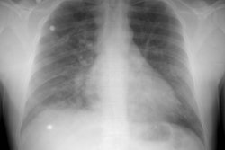
CXR demonstrates a unilateral ipsilateral alveolar filling pattern involving the entire lung (rarely only one lobe is affected) within a few hours of reexpansion of the lung. The edema may progress for 24 to 48 hours, and may persist for 4 to 5 days. The edema usually resolves in 5 to 7 days. Acutely, differential considerations would include a rapidly evolving pneumonitis in an immune compromised patient, aspiration pneumonia, and hypostasis in the dependent lung following prolonged decubitus positioning. Sometimes the edema can be bilateral, suggesting the release of mediators which affect the contralateral lung [2].REFERENCES:
(1) J Thorac Imag 1996; 11: p.198-209
(2) J Thorac Imag 1998; Ketai LH, Godwin JD. A new view of pulmonary edema and acute respiratory distress syndrome. 13: 147-171
(3) Radiographics 1999; Gluecker T, et al. Clinical and radiologic features of pulmonary edema. 19: 1507-1531


