Intramural hematoma with penetrating ulcer:
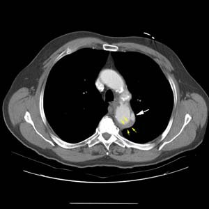
This elderly patient presented with severe chest pain radiating to the back. The CT scan revealed a penetrating aortic ulcer (white arrow) arising from the lateral aspect of the distal aortic arch. Smooth high density mural thickening of the aortic wall (yellow arrows) can be seen and is consistent with an intramural hematoma.
(Click image to enlarge)

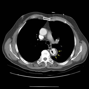
More inferiorly, there is displacement of an intimal calcification (black arrow) by the high density intramural hematoma (yellow arrows).
(Click image to enlarge)


