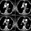
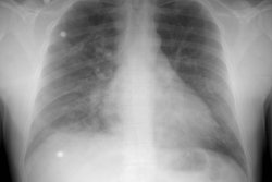
CT findings of pulmonary parenchyma in Takayasu arteritis.
Takahashi K, Honda M, Furuse M, Yanagisawa M, Saitoh K
PURPOSE: Our goal was to describe the pulmonary parenchymal manifestations of Takayasu arteritis visualized on CT. METHOD: We assessed the CT findings for the pulmonary parenchyma in 25 patients with Takayasu arteritis and compared them with those visualized by pulmonary angiography (n = 20) and radionuclide perfusion scintigraphy (n = 19). RESULTS: A review of the CT scans revealed a total of 33 low attenuation areas in the lung (11 patients), subpleural reticulolinear changes (12 patients), and pleural thickening (9 patients). The low attenuation areas were preferentially seen in patients with pulmonary arteritis and corresponded to pulmonary angiographic staining and scintigraphic perfusion defects. No significant correlation was found between other CT findings and pulmonary arteritis. CONCLUSION: The findings suggest that pulmonary low attenuation areas observed on CT represent regional hypoperfusion due to pulmonary arteritis. We speculate that pulmonary thromboembolism may contribute to other CT findings for the pleura and adjacent lung.


