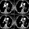
MR of a Coarctation of the Aorta
Image submitted by Dr. Scott D. Flamm, Cardiovascular MR Section, Cleveland Clinic Foundation


The Spin echo image on the left demonstrates a focal area of narrowing of the thoracic aorta just beyond the arch. Post-stenotic dilatation of the descending thoracic aorta is also evident. A cine gradient image nicely demonstrates a turbulent jet of flow passing through the area of narrowing which appears as an area of signal void on the image.


