Idiopathic Giant Bullous Emphysema: (Vanishing Lung Syndrome)
View cases of giant bullous emphysema
Clinical:
Vanishing lung syndrome has also been referred to as "type 1 bullous disease" or "primary bullous disease of the lung." It is characterized by the presence of large, predominantly upper lobe bullae. Although the disorder is always bilateral, the involvement is asymmetric with one side demonstrating more extensive disease. The disorder is most commonly observed in young men who are generally smokers, although it has also been described in non-smokers. Vanishing lung is a progressive disorder. The radiographic criteria necessary for the diagnosis is the presence of giant bullae in one or both upper lobes, occupying at least one-third of the hemithorax, and compressing surrounding normal lung. Infection of the bullae is rare. Spontaneous pneumothorax is more prevalent in patients with the disorder. However, it is extremely important not to mistake the bullae themselves for a pneumothorax. If treated with a thoracostomy tube, this will result in a large broncho-pleural fistula and lung collapse (because the tube is within the bulla, and not the pleural space). Palliative bullectomy can result in clinical improvement in symptomatic patients. HRCT is very useful in cases being considered for surgical resection as it may identify underlying severe centrilobular empysema which would preclude surgery (paraseptal emphysema does not preclude surgical resection). Preventive surgery for bullae occupying less than one-third of the hemithorax has not been shown to result in functional improvement. (See also Lung Reduction Surgery)
X-ray:
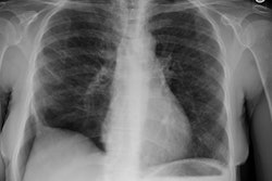
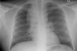
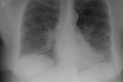
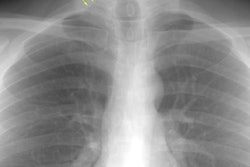
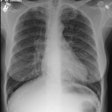
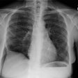
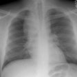
CXR: Plain films will demonstrate the presence of bilateral bullous emphysema with a predominantly upper lobe distribution. The bullae are generally between 2 to 8 cm in diameter.
Computed tomography: HRCT will not only demonstrate the presence of multiple large bullae, but has also be used to demonstrate the presence of paraseptal emphysematous change in all patients with this disorder. Although paraseptal emphysema is not usually symptomatic, it is believed that these peripheral air spaces coalesce to form the giant bulla which characterize this disorder. Centrilobular emphysema may also be identified in these patients and is only seen in smokers.
REFERENCES:
(1) AJR 1994; Feb 162 (2):279-282
(2) J Thorac Imag 1986; 1(2): 75-93
(3) AJR 2000; Waitches GM, et al. Usefulness of the double wall sign in detecting pneumothorax in patients with giant bullous emphysema. 174: 1765-1768
