Rounded atelectasis: Atypical appearance
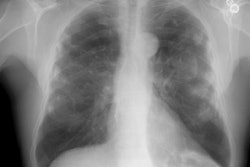
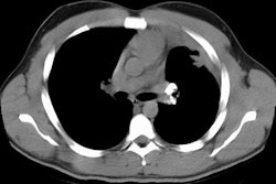
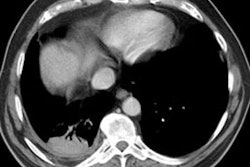
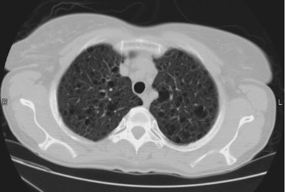
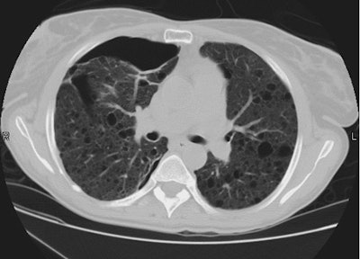
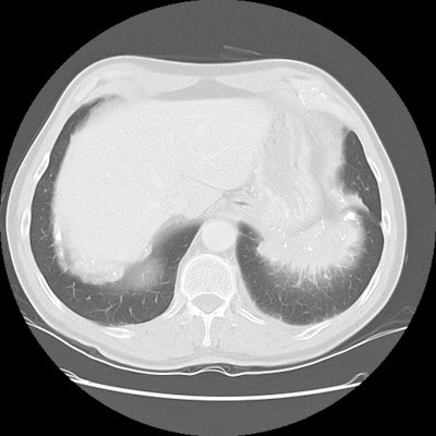
The patient shown on the CT images below presented for evaluation of an enlarging abnormality on CXR. The CT demonstrates exuberant pleural plaque disease along the right diaphragmatic surface. There is a soft tissue mass within the left lower lung that contains a large eccentric calcification. Lung markings radiate into this lesion from the adjacent lung parenchyma and there is distortion of the major fissure. The finding suggested rounded atelectasis, however, the lesion had more aggressive features more inferiorly (see lower CT images). PET images are shown below CT's.
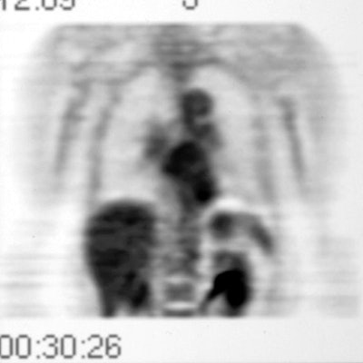
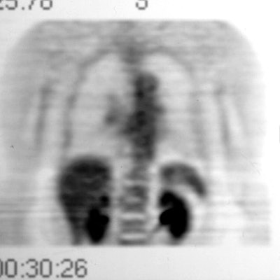
A PET-FDG scan was performed to determine if the lesion demonstrated increased metabolic activity. The lesion appeared as a region of decreased activity above the surface of the left hemidiaphragm. This finding suggested the lesion to be rounded atelectasis which has been shown to not be metabolically active on FDG-PET imaging. The patient had the lesion surgically resected and it was found to be rounded atelectasis and pleural plaque.
