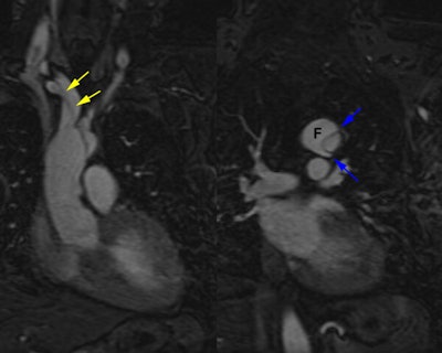
Type A dissection:
The MR exam was performed to better assess possible extension of the dissection into the great vessels. Off axis sagittal images demonstrated the dissection extending into the right inominate artery and right subclavian artery (yellow arrows). The smaller lumen is the true lumen. The "beak sign" described on CT as a feature can also be seen on these MR images. Small "beaks" of contrast in the false lumen (blue arrows) extend around the margins of the true lumen. |
|


