Amniotic Fluid Embolization:
Clinical:
Amniotic emboli are a rare phenomenon- occurring in 2 to 7.7 per
100,000 deliveries [3]. The condition is associated with very high
maternal (80% [2]) and fetal (40%) mortality. Most amniotic fluid
emboli develop during the first stage of labor and occurs when
amniotic fluid is forced into the bloodstream through small tears
in the uterine veins during labor [2].
Risk factors associated with the development of amniotic fluid emboli include advanced maternal age, multiparity, prolonged gestation, fetal demise, use of uterine stimulants/labor induction, large fetal size, premature rupture of the membranes, instrumented vaginal delivery, cesarean section, pregnancy-induced hypertension, polyhydramnios, placenta previa, prior cervical trauma, and meconium staining of the amniotic fluid [1,3,4]. The amniotic fluid contains substances such as leukotrienes and prostaglandins which produce a pulmonary vasospasm. Subsequently there is severe pulmonary hypotension and marked hypoxia which results in the death of 50% of patients within the first hour (most deaths occur within 2 hours of the onset of symptoms).
The classic clinical presentation is abrupt dyspnea, cyanosis, and shock [2]. This is followed (within minutes) by cardiorespiratory arrest and severe pulmonary edema. The symptoms begin during spontaneous labor in about 70% of patients and following parturition in the remaining 30% (10% with spontanoeus delivery and 20% with c-section) [2]. A severe consumptive coagulopathy (disseminated intravascular coagulation) occurs in 40% of patients. Convulsions are also common.
X-ray:
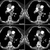
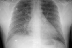
CXR/CT demonstrates a pulmonary edema/adult respiratory distress syndrome appearance with diffuse bilateral homogeneous/heterogeneous opacities. Thickening of the interlobular septa may also been seen [3].
REFERENCES:
(1) AJR 2000; Rossi SE, et al. Nonthrombotic pulmonary emboli. 174: 1499-1508
(2) Radiographics 2003; Han D, et al. Thrombotic and
nonthrombotic pulmonary arterial embolism: spectrum of imaging
findings. 23: 1521-1539
(3) AJR 2017; Unal E, et al. Nonthrombotic pulmonary artery
embolism: imaging findings and review of the literature. 208:
505-516
(4) Radiographics 2017; Plowman RS, et al. Imaging of
pregnancy-related vascular complications. 37: 1270-1289
(5) Radiographics 2020; Gonzalo-Carballes M, et al. A pictoral review of postpartum complications. 40: 2117-2141


