CT Features to Distinguish the True from False Channel:
1- The beak sign: Small "beaks" of contrast or thrombus can usually be identified at the lateral margins of the false lumen [6]. 2- Cobwebs: Cobwebs are ribbons of media which are incompletely sheared off by the dissection and are located exclusively in the false lumen [6]. 3- Outer wall calcification: Although outer wall calcifications may be seen in the false lumen of a chronic dissection, they are generally not seen in the outer wall of the false lumen of an acute dissection (ie: outer wall calcifications imply the true lumen) [6]. 4- Eccentric calcifications along the dissection flap: Calcifications along the dissection flap are usually located along the true lumen side of the flap [6]. 5- Intralumenal thrombus: Intralumenal thrombus is more commonly seen in the false lumen of both acute and chronic dissections, although in patients with aortic aneurysms thrombus may be present in the true lumen as well [6]. 6- Larger lumen: The larger lumen is usually the false lumen. The false lumen will often compress the true lumen as well. [6]. 7- Wrap around: In patients with involvement of the aortic arch, it is common for one lumen to appear to wrap around the other lumen (sometimes giving the appearance of three lumens). In such cases, it is the false lumen that wraps around the true lumen [6].
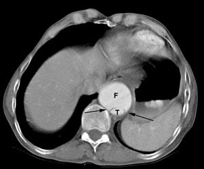
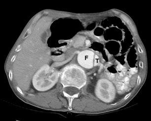
The "beak sign" is demonstrated by the image on the left. In this acute dissection small beaks of contrast (black arrows) extend from the false lumen (F) around the margins of the true lumen (T). The false lumen (F) is also commonly larger than the true lumen (T). |
|
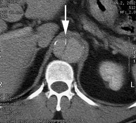
Eccentric calcifications along the dissection flap: Calcifications along the dissection flap are usually located along the true lumen side of the flap (white arrow). In this case the true lumen is to the right and the false lumen to the left. |
|
