It's said that "Dead men tell no tales," but a 4,000-year-old female mummy that was recently re-examined with a 64-slice CT scanner revealed many of her secrets to paleoimaging experts.
Researchers from Quinnipiac University in Hamden, CT, used 64-slice CT on the Pa-Ib (pronounced pie-eeeb) mummy to identify at least five cloth bundles, containing her bodily organs, in the thoracic and abdominal regions.
"We got some incredible images," noted Gerald Conlogue, professor of diagnostic imaging at Quinnipiac's School of Health Sciences, adding that some of the 3D software was used in the "Avatar" science fiction film.
Repeat imaging
Pa-Ib was originally scanned in 2006 with an eight-slice scanner, and the goal of the scans at that time was to verify whether it was an authentic Egyptian mummy. Pa-Ib is one of the big attractions at the Barnum Museum in Bridgeport, CT, and was reportedly acquired by P.T. Barnum's wife through the Egyptian Consulate in the 1890s. The scans verified that Pa-Ib was indeed a real mummy and not one of Barnum's elaborate hoaxes.
This time, Conlogue and colleagues decided to use a 64-slice system (Aquilion 64, Toshiba America Medical Systems, Tustin, CA) as well as 3D software to gain additional insight into what Pa-Ib's life might have been like in ancient Egypt. The scans were conducted in January.
In addition, Ronald Beckett, Ph.D., a professor of respiratory care and chairman of cardiopulmonary sciences and diagnostic imaging at Quinnipiac, used an endoscope camera to probe the mummy's skull. He inserted the endoscope into Pa-Ib's right nostril, tracing the path that the Egyptians had made to remove her brain.
After Pa-Ib's death, a hook was pushed through her nose and then used to punch through the orbital bone in her eye socket into her brain cavity. Her brain was then scrambled, and she was turned over so her brain came out through her nose. The cavity was then filled with resin, which shows on the scans.
The Egyptian translation for brain is "snot maker," laughed Conlogue in an interview with AuntMinnie.com.
"You can see the grooves that indicate the shape of the tool that was used; it looks like it had kind of a scoop on it," explained Conlogue. "It was either a rush job or the people doing it weren't very skilled, because instead of going through the thin bone at the top of the skull, they went through her nostril and broke through the bone that forms the medial wall orbit."
 |
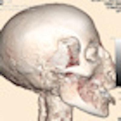
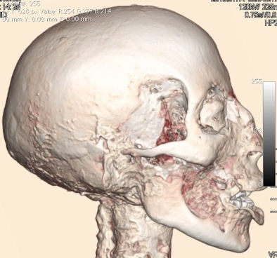
| This 3D surface reconstruction shows the mummy's curled lip, exposing her teeth; a bit of textile wrapping covering her eye, resembling an eye patch; and a red area of dried muscle on her cheek. All images courtesy of Gerald Conlogue. |
The mummy's worn-down teeth seemed to be in the worst shape, showing she probably had sand in her food and wasn't from the upper classes.
"Because she lacks a lot of degenerative changes in her skeleton, this is probably someone who wasn't doing a lot of repetitive motion," Conlogue said. "She probably didn't do a lot of manual labor. It's more than likely she died of an acute illness like a respiratory infection."
Beckett and Conlogue participated in the original 2006 examination of Pa-Ib. They are the founders and executive directors of Quinnipiac's Bioanthropology Research Institute and hosts of "The Mummy Road Show," a TV show that aired on the National Geographic Channel.
The calcium buildup in one bundle suggests either an infection or a salt residue, which was used to preserve the organs, Conlogue said. Dr. Ramon Gonzalez, director of the radiology assistant program at Quinnipiac and an assistant clinical professor of radiology at Yale University in New Haven, CT, determined Pa-Ib was younger than 40 years when she died.
"She had remarkably well-preserved joints, the ossification of her bones was very good, and there was no evidence of trauma or fractures," Gonzalez told AuntMinnie.com. "She was a very healthy individual."
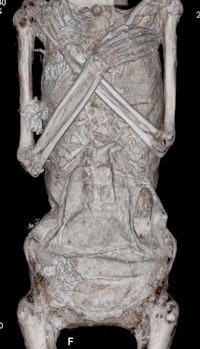
Using 3D software, they were able to see the area in the chest where those who prepared Pa-Ib's body made a large incision to the abdomen to remove the viscera, Gonzalez said.
 |
| This 3D reconstruction shows the incision made on Pa-Ib's left side [hole above her hip on right side of image], which was made during the mummification process to remove her organs. |
Some of the images distinctly show the woven pattern of the cotton used to wrap the organs.
"We could clearly see the texture of the burlap that was used to wrap the packets; that was really amazing," Gonzalez said. "You can see the elongated, cylindrical shape because they had to introduce it back into the cavity through the incision, which was a really small orifice."
Another interesting thing, noted Gonzalez, was how little evidence there was of arthritic change, but he did find one osteophyte on her dorsal spine.
"Some remains of her skin were still attached to her legs, and you can see how well the feet were wrapped," Gonzalez said. "You could tell she was young: her feet were still pristine; there was no evidence of arthritis or heel spurs."
The images showed Schmorl's nodes on the superior surface of the T12, L1, and L4 vertebral bodies, Conlogue said. He also noted that Pa-Ib's knees were free of degenerative changes and the anterior and posterior cruciates were well defined and normal in appearance.
Researchers had previously thought Pa-Ib might have given birth because the 2006 exam showed evidence of arthritis in the pelvic area. But Conlogue said malalignment of the symphysis pubis was due to postmortem migration of the pelvic ligament. "Since there was a lack of sclerotic or hypertrophic changes at the pubic symphysis, there appeared to be little evidence of a pregnancy," he explained.
The mummy's wrappings were put on tightly, which could have changed the position of bones, or the bone displacement could be a result of the bones drying and shriveling, Conlogue said.
The mummy also underwent an MRI scan, but the imaging resulted in no additional findings, due to the artifact's dry condition.
"It's very exciting because we can share these images with people around the world," Conlogue said.
"What makes these mummies so fantastic is this is an actual person who walked the earth about 3,500 years ago. That's pretty amazing," Conlogue observed. "Within her body are all the secrets to her life. A person's life history is really recorded in the bones, so with a 64-slice scanner we can coax those secrets out of her. It's like a book with 4,000 pages, and we're looking for typos on the page."
By Donna Domino
AuntMinnie.com contributing writer
February 11, 2010
Related Reading
Mummy CT scans show ancient Egyptians had atherosclerosis, November 18, 2009
Austrian study uses MDCT to uncover tale of 12 mummies, October 5, 2009
CT reveals Nefertiti's secrets, April 1, 2009
Philips CT images Egyptian mummy, February 5, 2009
Imaging reveals truth about Barnum mummy, October 31, 2006
Copyright © 2010 AuntMinnie.com


