Read prone virtual colonoscopy dataset, check. Read supine dataset, check. Practitioners know that examining both -- and effectively double reading every virtual colonoscopy exam -- is necessary to establish that findings on one dataset are present on the other.
But a new endoluminal surface registration approach has succeeded in cobbling together the two datasets more completely than previous methods to create a virtual endoluminal view similar to that of conventional colonoscopy, according to a study in Medical Physics.
The method works despite divergent twists and turns in the two datasets, and it can even be applied to regions of local colonic collapse by simply ignoring the collapsed segments, say researchers from University College London.
"Our method simplifies the problem of aligning the prone and supine surfaces from a 3D to a 2D task. This is achieved by mapping the full endoluminal surface to a cylindrical parameterization using a conformal mapping," explained lead author Holger Roth, PhD, along with Jamie McClelland, PhD, and colleagues (Medical Physics, June 2011, Vol. 38:6, pp. 3077-3089).
Radiologists spend considerable time visually matching up endoluminal locations between prone and supine virtual colonoscopy (also known as CT colonography or CTC) images to determine whether detected polyps are real or residual fecal matter in the colon. The colon can be considerably deformed as the patient changes position, and severe underdistention can lead to colonic collapse.
"This complicates the interpretation task; identifying corresponding regions of endoluminal surface between prone and supine acquisitions is difficult, prolongs reporting time, and may lead to errors of interpretation," the group wrote.
A reliable method for registering prone and supine data could improve accuracy and cut interpretation time, and the technique could also be incorporated into computer-aided detection (CAD) systems to improve the accuracy of CAD, the authors wrote.
Roth and his team proposed a method of automatically establishing spatial correspondence between prone and supine endoluminal surfaces of the colon after surface parameterization.
Cylindrical parameterization
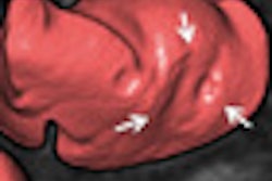
Conformal maps have been used by several groups to "virtually dissect" the colon and produce flattened 2D images of the endoluminal surface -- techniques that were developed to speed interpretation, the authors explained. Typically applied to surface mesh triangulations to simplify the representation of a 3D object, they are used to map 3D surfaces to a 2D space while preserving local angles. Those surfaces, or S, can be parameterized using a discrete conformal mapping method, which maps the entire surface while preserving the appearance of local structures such as polyps and haustral folds.
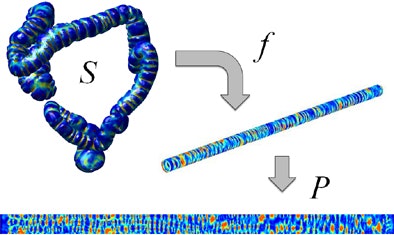 |
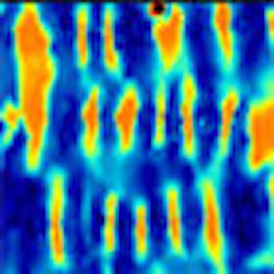
| The image above depicts the principle of registration between endoluminal surfaces S of the colon derived from prone and supine CT colonography using cylindrical 2D parameterizations P. The color scale indicates curvature-based measurements on the 3D surfaces, here shape index. These measurements can be used to compute the transformation between the two cylinders using nonrigid registration. All images courtesy of Holger Roth, PhD. |
To extract the endoluminal colonic surface S, the inflated lumen L is segmented using the method described by Slabaugh et al, the group wrote. The endoluminal colonic surfaces are then modeled as triangulated meshes on the gas-tissue border in the CTC images.
Curvature information from the original 3D surfaces is used to determine correspondence. A nonrigid registration method is used to align the local structures accurately.
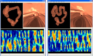 |
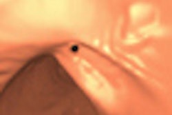
| Endoluminal view of a polyp after using the proposed registration method to find the correspondence between the prone and supine colon surfaces, indicated by the black dot in both 3D renderings and 2D representations. |
Results
A training dataset was used to find suitable parameters to constrain the cylindrical registration method. Those same registration parameters were then applied to a different set of 13 validation cases that comprised eight fully distended cases and five cases exhibiting multiple colonic collapses.
All polyps were found to be well aligned, with a mean registration error of 5.7 ± 3.4 mm. An additional 1,175 reference points on haustral folds spread over the entire endoluminal colon surface resulted in an error of 7.7 ± 7.4 mm. Overall, 82% of folds were aligned correctly after registration, with a further 15% misregistered by just one fold, the authors reported.
The method is based on the assumption that the colon can undergo large deformations when the patient changes position, but that the local shape of surface structures, such as haustral folds, remains similar enough to align the surfaces of two scans.
That the method relies on extracted colonic surfaces of high quality for robust segmentation is a "significant impediment to the clinical adoption of our method," they wrote, because flawed acquisitions remain common in CTC despite improvements in recent years.
Future work, they added, will also address cases of widely varying distentions between prone and supine views, insufficient tagging, and finding automated methods of addressing local underdistention with more complex collapses.


