Conebeam CT (CBCT) helped clinicians remove an ectopic third molar and an infected cyst from a patient's maxillary sinus, which is a rare site for a tooth to erupt, according to a recent case report published on the open-access platform F1000Research.
The case showcases the importance of using the appropriate tools to evaluate patients and to prepare clinicians for procedures, according to the authors.
"The CBCT assessment of our case allowed us to precisely locate the ectopic molar and showed the exact extension and effect of the associated pericoronal lesion on the surrounding structures," wrote the group, led by Khairy Elmorsy, DMD, of the oral and maxillofacial surgery department at Cairo University (F1000Res, April 16, 2020).
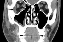
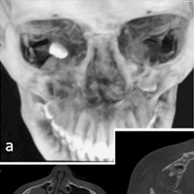
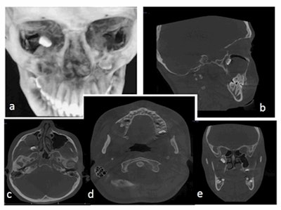 Multiplanar CBCT sections: (a) three-dimensional, (b) sagittal, (c, d) axial, and (e) coronal slices showing the posterosuperior position of the ectopic molar inside the maxillary sinus with associated lesion that surrounds the crown of the tooth. All images courtesy of Khairy Elmorsy, DMD, et al. Licensed under CC BY-NC 4.0.
The development of ectopic teeth in nondental areas, specifically the maxillary sinus, is rare. A review of scientific journals revealed reports of only 51 patients having ectopic teeth in the maxillary sinus. Third molars represented the higher prevalence of ectopic teeth, accounting for 21 of the cases, the authors wrote.
Though the cause of ectopic eruptions is unclear, some are the result of a developmental condition, such as cleft palate, or a pathological process, such as the development of large cysts, that can push tooth buds to unusual places. Though panoramic x-rays are routinely used to examine this type of eruption, CBCT helps better localize the ectopic tooth and assess any associated lesions before a patient undergoes surgery.
A bad taste and a foul odor
In the current case, a 13-year-old girl visited an oral and maxillofacial surgery clinic after she experienced discharge, which had a nasty taste and foul smell, that oozed from her upper right second molar for about two weeks, according to Elmorsy and colleagues.
Oral surgeons examined her and found no caries or internal or external swelling. However, a panoramic x-ray showed an ectopic eruption of her right maxillary third molar in her maxillary sinus. A hyperdense lesion surrounded her molar's crown, the authors wrote.
To get a better view, the patient underwent a CBCT scan. The scan showed that the teenager's ectopic third molar had incompletely formed roots located in the posterosuperior aspect of the right maxillary sinus with a close approximation to the orbital floor superiorly and pterygoid plates posteriorly. The lesion measured 23 × 36 × 35 mm, occupied almost the entire cavity of her right maxillary sinus, caused mediolateral expansion of the alveolar ridge, and encapsulated the tooth. The scan also showed how the lesion was obliterating the right maxillary sinus floor and buccal cortical plate. This explained why the girl was experiencing discharge, the authors noted.
Treatment strategy
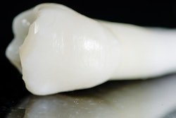
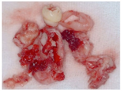 The removed underdeveloped ectopic molar and cystic lining.
The patient underwent a lateral sinus antrostomy, which involves entering the maxillary sinus via the canine fossa, based on the exam and imaging results. The surgeon drained pus from the cystic lining and then removed the cyst, its lining, and the ectopic molar. The patient was prescribed antibiotics and acetaminophen and was given one dose of steroids to prevent her cheek from swelling.
She recovered without further incident. During a three-month follow-up visit, she underwent another CBCT scan that showed some bone formation in the mediolateral dimension, which indicated that the bone had started to heal.
A valuable resource
Enhanced imaging such as CBCT is becoming an increasingly important tool for dentists. Scans aid dentists in diagnosing patients and developing treatment plans, according to the authors.
"[The CBCT assessment] allowed the preoperative identification of the cause of the purulent discharge and aided the surgeon during the surgical procedure, mainly to avoid any surgical complications due to the proximity to the orbital floor and pterygoid plates," they wrote.


