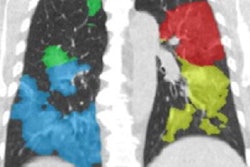
A disease associated with COVID-19 that causes multiple organ failure in children presents imaging findings that are different than a typical acute infection, indicating that COVID-19 is likely far more than a respiratory illness in kids, according to an article published online July 30 in Radiology: Cardiothoracic Imaging.
Based on their experience with 20 recent cases of multisystem inflammatory syndrome in children (MIS-C), a multi-institutional research team led by Dr. Abbey Winant of Boston Children's Hospital said that MIS-C is likely a postviral hyperinflammatory process that is known to cause multiorgan damage, such as heart disease, liver injury, kidney failure, and gastrointestinal and dermatologic manifestations.
"This signals an important paradigm shift in our understanding of pediatric COVID-19 infection: from a primarily respiratory illness to multiorgan system disease," the authors wrote.
Three main thoracic imaging findings -- heart failure, acute respiratory distress syndrome (ARDS), and pulmonary embolism (PE) -- have been observed in patients with MIS-C, according to Winant and colleagues from Boston Children's Hospital and the Children's Hospital at Montefiore and Montefiore Medical Center in New York City.
These results, as well as other emerging extrathoracic imaging findings, are different than the typical findings found in acute COVID-19 cases in children (see table below).
| Typical COVID-19 imaging findings vs. MIS-C findings in children | ||
| Typical acute COVID-19 case | MIS-C associated with COVID-19 | |
| Pulmonary findings | Bilateral, lower lobe predominant peripheral/subpleural ground-glass opacities and/or consolidations; halo sign (early phase) | Pulmonary edema;
ARDS, possibly asymmetric |
| Pleural findings | None known at time of publication | Pleural effusions |
| Cardiovascular findings | None known at time of publication | Heart failure/left ventricular systolic dysfunction, pericardial effusion, PE (segmental PE has been observed so far), coronary artery dilatation |
| Extrathoracic findings | None known at time of publication | Mesenteric lymphadenopathy, hepatomegaly, gallbladder wall thickening, echogenic renal parenchyma, ascites |
"As previously described, it has been suggested that MIS-C associated with COVID-19 and late-stage adult COVID-19 may be characterized by a similar hyperinflammatory milieu, which may account for some overlap in imaging findings and pathophysiology, although more scientific evidence is needed for validation," Winant and colleagues wrote.
More scientific evidence is also necessary to guide clinical and imaging study decisions in these patients, according to the authors. Based on their preliminary observations of the condition in their patients, however, the researchers said that a judicious approach to imaging pediatric patients with this syndrome may need to include echocardiography, abdominal imaging, and CT pulmonary angiography in those with a high clinical suspicion for pulmonary embolism, in addition to typical chest radiographs and/or CT.
"As knowledge and scientific evidence about the imaging findings of MIS-C associated with COVID-19 grow, better understanding of the characteristic imaging findings and the need for specific imaging evaluations is expected in the future," the authors wrote. "In addition, because there is currently a lack of pathologic data explaining the underlying causes and imaging findings of MIS-C, future studies focusing on the radiology-pathology correlation will shed light on this new and challenging disorder, unique to the pediatric population."


