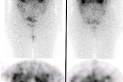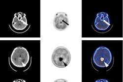
CHICAGO - The combination of gallium-68 (Ga-68) DOTATATE PET and MRI can greatly improve the assessments of how well patients with meningiomas respond to surgery and/or radiation treatment, according to a study presented Sunday at the RSNA 2019 meeting.
The dynamic hybrid modality was instrumental in the accurate assessment of 61% of the malignant recurrent and progressive lesions, improved the characterization on extent of disease, and confirmed parenchymal and osseous invasion in a disease that carries with it a poor prognosis.
Meningiomas are the most common primary intracranial tumors, with total resection often the course of treatment, although it can only be accomplished in 50% to 66% of cases, according to previous research. Patients also face a greater risk of disease progression and reduced progression-free and overall survival.
The numbers are "surprisingly high," said Dr. Jana Ivanidze, PhD, assistant professor in the department of diagnostic radiology at Weill Cornell Medicine, who presented the findings, "given that we think of meningiomas as this slow-growing lesion that we have nothing to worry about; but progression-free survival at six months is only 30%, even in grade 1 or grade 2 cases."
Contrast-enhanced MRI is the gold standard for diagnosis and treatment planning. However, MRI can have limited accuracy in distinguishing recurrence from treatment effect in the postsurgical and postradiation setting. Ga-68 DOTATATE, on the other hand, is a PET radiotracer that targets somatostatin receptor 2 (SSTR2) with high affinity. This attraction is particularly important because SSTR2 is expressed at a high level in meningiomas.
Thus Ivanidze and colleagues sought to evaluate Ga-68 DOTATATE PET/MRI in a prospective clinical cohort of patients with meningioma.
For this study, the researchers analyzed 18 patients with clinically suspected or pathology-proven meningioma who were imaged over a period of six months. Ga-68 DOTATATE PET/MRI was acquired in 3D list mode over 50 minutes, beginning five to 15 minutes after injection. Among the data gathered were maximum standard uptake values in meningiomas and suspected post-treatment changes, using the pituitary gland as a positive reference and superior sagittal sinus as the background reference.
The researchers found Ga-68 DOTATATE PET showed unique uptake patterns for meningiomas, pituitary glands, and post-treatment changes across all subjects. The modality also improved the researchers' ability to visualize the extent of disease and confirm parenchymal and osseous invasion.
As a result, the modality detected 31 unique lesions from the scans, which included 19 confirmed meningiomas (61%) and seven lesions (23%) with low avidity. The latter finding would suggest a change in the disease status of the patient after treatment. The remaining five lesions (16%) could not be definitively diagnosed and were deemed indeterminate.
Given the results, the researchers concluded that Ga-68 DOTATATE PET/MRI is a "promising tool" to help clinicians assess meningiomas after treatment and allowed for "improved diagnosis and extent-of-disease evaluation without increasing acquisition time."
The findings also serve as the basis for the researchers' prospective DOMINO-START clinical trial, which recently opened to enrollment. This study will include patients who undergo resection for somatostatin receptor-positive central nervous system tumors, focusing on meningioma and including esthesioneuroblastoma, hemangioblastoma, medulloblastoma, paraganglioma, pituitary adenoma, and SSTR-positive systemic cancers metastatic to the brain, such as small cell carcinoma of the lung.
They also plan to advance the current Ga-68 DOTATATE PET/MRI research to see how the dynamic hybrid modality might help assess the response of patients to meningioma radiation treatment planning, as well as positive tumors of the head, brain, and neck.
Study disclosures
Advanced Accelerator Applications, a Novartis company, provided a research grant for this study.


