Flortaucipir-PET and blood tests have correlated two key biomarkers of chronic traumatic encephalopathy (CTE) in military personnel who experienced traumatic brain injuries (TBI) from blasts, according to a study published online on February 25 in Molecular Psychiatry.
Researchers from Mount Sinai and the James J. Peters Veterans Affairs (VA) Medical Center observed elevated levels of tau tangles and a certain blood chemical associated with TBI, both of which could be indications of current or future acute brain degeneration and possible progression to CTE as a result of brain injury from an improvised explosive device (IED).
"These findings have implications for understanding the relationship of chronic blast-related injury to human neurodegenerative diseases including CTE," wrote the authors, led by Dara L. Dickstein, PhD, adjunct assistant professor of neuroscience, and Rita De Gasperi, PhD, associate professor of psychiatry, both from the Icahn School of Medicine at Mount Sinai. "The current data provide evidence ... to support the existence of a relationship involving blast injury and clinical neuropsychiatric syndromes."
Estimates are that 10% to 20% of veterans who served in Iraq and Afghanistan sustained mild TBI from IEDs and other explosions. While symptoms from mild TBI dissipate within months of an incident, neurological deterioration and behavioral dysfunction can linger for years in other many personnel.
Two distinct chemical changes occur during neurodegeneration. One action is the accumulation of tau neurofibrillary tangles, which are associated with the onset of dementia, Alzheimer's disease, and CTE. Through PET imaging, flortaucipir (Avid Radiopharmaceuticals) has shown its ability to bind to tau neurofibrillary tangles in postmortem brain tissue of patients with Alzheimer's disease.
A second chemical sign is the leakage of a protein known as neurofilament protein-light chain (NfL) from the brain into the blood. Elevated levels of NfL are evident in patients with a variety of brain injuries, including mild TBI and neurodegenerative diseases.
To explore a connection among neurodegeneration, TBI, and these potentially destructive proteins, the researchers analyzed 10 veterans (mean age, 41.2 ± 9.4 years) who experienced between one and 50 or more blasts, had at least one TBI, and reported chronic behavioral issues and cognitive deficiencies. The cohort also included seven healthy age-matched subjects. All 17 participants underwent 3-tesla structural MRI scans (Biograph MR, Siemens Healthineers), followed by PET imaging (Biograph mCT, Siemens) with 370 MBq (10 mCi) of flortaucipir.
The researchers also targeted six cortical brain regions -- the frontal, temporal, parietal, precuneus, anterior cingulate, and posterior cingulate cortex -- where tau protein is known to accumulate after a traumatic brain injury.
Levels of NfL were assessed using an ultrasensitive single-molecule array (Simoa, Quanterix), a digital form of an enzyme-linked immunosorbent assay (ELISA) that allows detection of proteins at subfemtomolar concentrations.
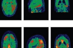
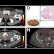
The PET images revealed that five (50%) of the 10 veterans had excessive retention of flortaucipir at the white/gray-matter junction in the frontal, parietal, and temporal brain regions, which indicates tau accumulation and the likelihood of neurodegeneration by eventually atrophying brain cells in those areas. The researchers also observed elevated levels of NfL in the plasma of veterans displaying excess flortaucipir retention. As with tau accumulation, evidence of NfL could be an indication of neurodegenerative decline.
"While the amount of [flortaucipir] retention varied, the patterns of retention consistently resembled the pathognomonic distribution of CTE tauopathy, with foci of [flortaucipir] retention located deep in the sulci at the white/gray matter junctions in frontal, parietal, and occipital brain regions," the authors wrote.
While the findings are not conclusive, Dickstein, Gasperi, and colleagues added that the findings "support an emerging consensus" that NfL and other biomarkers could play an important role in monitoring postconcussive recovery and do warrant additional research.


