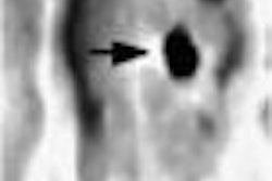
SEATTLE - For patients with "bulky" enlarged cervical lymph nodes, CT fusion with 18fluorodeoxyglucose (FDG) PET improves spatial allocation and the visualization of pathology, according to an Austrian study presented yesterday at the American Roentgen Ray Society meeting.
Dr. Gottfried Schaffler, a radiologist at the University Hospital in Graz, Austria, and the University of California, San Francisco, discussed the results of a study that evaluated the potential clinical value of digital image fusion of spiral CT and FDG-PET in patients with suspected cervical malignancies.
The study looked at 22 patients, including 12 men and 10 women (mean age, 49) who had undergone spiral CT scanning for primary staging of malignancies including lymphoma, melanoma, and lung cancer. Most of the patients suffered from lymphoma. The mean lag time between spiral CT and PET scanning was 18 days, Schaffler said.
Patients underwent CT scanning of the neck and craniocaudal region using a Somatom Plus IV scanner (Siemens Medical Solutions, Iselin, NJ) using a slice thickness of 3 mm following bolus injection of 100 ml of a nonionic contrast agent.
All patients subsequently underwent FDG-PET imaging one hour following injection of 300-375 MBq of 18fluorodeoxyglucose, using a high-resolution Siemens ECAT HR PET scanner.
A computer workstation was used to fuse the image data sets using Hermes software (Nuclear Diagnostics, Haegersten, Sweden) which provided automatic adaptation of pixel sizes and semiautomatic adaptation of different body axes using anatomical landmarks, Schaffler said. Average fusion time was about 11 minutes.
"The digital images were reconstructed and displayed in sagittal, coronal, and axial planes," he said. "All images were reviewed and evaluated by consensus of two radiologists, and also the digital images were reviewed by consensus."
Schaffler and colleagues found 42 pathologically enlarged cervical lymph nodes in 4 patients. FDG-PET showed homogeneous and pathologically increased radiotracer uptake in all 42 enlarged lymph nodes. FDG-PET improved the spatial allocation in 32 of 42 pathologically enlarged lymph nodes found in two patients presenting with "bulky" disease.
"Digital image fusion improved the spatial allocation in these [two] patients, because on the PET scan we found heterogeneous [FDG uptake], and on the image fusion we could align them to the enlarged cerebral lymph nodes," Schaffler said. "In the remaining two patients, we found 10 moderately enlarged, randomly distributed and separated lymph nodes with homogeneous[ly] increased FDG uptake, and these patients did not [benefit] from digital image fusion."
In summary, Schaffler said the study proved the technical feasibility of using digital image fusion to evaluate patients with pathologically enlarged cervical lymph nodes. Moreover, the commercially available fusion software proved more than adequate for the task. Fusion improved spatial allocation in patients presenting with "bulky" disease, but not in those with moderately enlarged nodes, and did not detect additional enlarged nodes, he said.
An audience member asked why it was necessary to perform an expensive PET procedure in patients who were presumably already undergoing therapy for lymphoma based on CT results. Schaffler said that having detailed information about the location of tumor foci within the enlarged nodes was potentially very useful for therapeutic procedures such as fine-needle biopsy.
By Eric Barnes
AuntMinnie.com staff writer
May 3, 2001
Click here to post your comments about this story. Please include the headline of the article in your message.
Copyright © 2001 AuntMinnie.com


