FDG-PET/CT is superior to CT for detecting primary and nodal disease in anal carcinoma and is "highly predictive" of increased progression-free survival, according to a presentation at the recent American Society for Radiation Oncology (ASTRO) meeting in Chicago.
Researchers from Southern California Permanente Medical Group in Los Angeles also found that CT alone failed to detect PET-positive lymph nodes in approximately 20% of patients.
"Our study helps confirm the superiority of FDG-PET scans to CT scans in visualizing primary and nodal disease in anal carcinoma," said lead author Dr. William Lien from the facility's department of radiation oncology. "Accurate staging is crucial to appropriate management of anal carcinoma, and FDG-PET scans should be obtained in all patients prior to initiation of definitive chemoradiation."
Patient conditions
From 2005 to 2008, 42 patients were treated in the department of radiation oncology for biopsy-proven, nonmetastatic anal carcinoma. Thirty-nine patients underwent pre- and post-treatment FDG-PET/CT scans. There were 26 women and 13 men, ranging in age from 37 to 91 years, with a median age at diagnosis of 60 years.
Among the cases, three patients were HIV positive, 32 patients were diagnosed with squamous cell carcinoma, five patients had basaloid carcinoma, and one patient was found to have adenosquamous carcinoma.
Two patients were treated with radiation alone, and 37 patients received a combination of chemotherapy and radiation. Thirty-six patients were treated only with external-beam radiation, and three patients received a combination of external-beam radiation and interstitial low-dose-rate implant brachytherapy with iridium-192 seeds.
Imaging protocol
All patients underwent FDG-PET/CT with a hybrid PET/CT scanner. CT images were obtained at 3.75-mm slice intervals without administration of intravenous contrast. Scans began at the base of the skull and extended through the proximal thighs.
PET images were obtained over the same region 40 to 60 minutes after the administration of FDG, with imaging times of three to six minutes per bed position. FDG doses ranged from 15 to 23 mCi, with a mean dose of 16.9 mCi.
All patients underwent FDG-PET/CT scans prior to any chemotherapy or radiation. Diagnostic CT scans of the abdomen and pelvis with intravenous contrast were also obtained and compared to FDG-PET/CT scans.
Post-treatment FDG-PET/CT scans were obtained, on average, 12.6 ± 6.8 weeks after the final day of radiation treatment, raging from one to 40 weeks. Progression-free survival was calculated from when the diagnosis was made until the patient's most recent follow-up or until the time of disease progression.
Two patients died during the course of the study. The researchers determined that one death was caused by metastatic anal carcinoma; the other death was due to metastatic renal cell carcinoma. Of the 37 remaining patients, six patients (15%) had disease progression, and two patients underwent abdominoperineal resection and are free of disease. Four other patients remain alive with cancer.
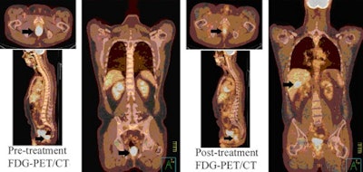 |
| Image shows an HIV-positive 41-year-old male with squamous cell carcinoma of the anus. Pretreatment FDG-PET/CT (left) demonstrated increased uptake in the anal canal (arrows) and a left internal iliac lymph node. Post-treatment FDG-PET/CT (right) demonstrated persistent uptake in the anal canal and new activity in the right lobe of the liver (arrows). The patient had biopsy-proven local and distant disease and died from local and metastatic disease 12 months after completion of chemoradiation. All images courtesy of Dr. William Lien. |
In their analysis, the researchers found that FDG-PET/CT detected the primary tumor in 38 of 39 cases (97%) in pretreatment staging, whereas CT alone detected primary tumors in 22 of 39 patients (56%).
In the study's post-treatment and progression-free-survival review, complete metabolic response was found in 26 patients (67%). Partial response was noted in 13 of 39 patients (33%). The two-year progression-free-survival rate for patients with complete metabolic response was 94.4%. For patients with partial response on post-treatment FDG-PET/CT scans, six patients developed progression of disease, with a two-year progression-free survival of 27.5%.
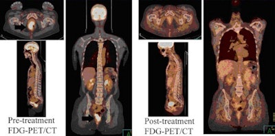 |
| Image shows a 47-year-old female with squamous cell carcinoma of the anus. Pretreatment FDG-PET/CT (left) demonstrated increased uptake in the right anal canal and two 5-mm perirectal lymph nodes. Post-treatment FDG-PET/CT (right) showed complete metabolic response. She remains without evidence of disease 31 months after chemoradiation. |
The researchers also repeated post-treatment PET/CT scans on four patients with a partial metabolic response. The clinical exams found that the tumors regressed in all four patients, who have had no evidence of disease on follow-up.
Based on the results, the researchers concluded that FDG-PET/CT is superior to CT in detecting primary and nodal disease in anal carcinoma and "post-treatment FDG-PET was highly predictive of progression-free survival and can be a useful tool in the post-treatment surveillance of patients treated with chemoradiation," Lien said. "Longer follow-up is necessary to determine whether or not this translates into an overall survival difference."
While optimal timing of post-treatment scans has not been determined, Lien said he and his colleagues "recommend obtaining FDG-PET scans eight to 12 weeks after completion of radiation."
By Wayne Forrest
AuntMinnie.com staff writer
November 25, 2009
Related Reading
PET/CT directs head and neck cancer treatment, November 17, 2009
FDG-PET/CT helps manage treatment for Crohn's disease, November 16, 2009
FDG-PET/CT helps assess pulmonary lymphangitic carcinomatosis, November 3, 2009
FDG-PET faces off against MDCT for gallbladder carcinoma, November 3, 2009
FDG-PET uptake helps predict vascular events in cancer patients, October 9, 2009
Copyright © 2009 AuntMinnie.com


