After remaining relatively stagnant for more than 50 years, the basic design of SPECT cameras has taken a quantum leap: Innovative developers have brought hardware and software improvements to the technology to reduce scan time and allow users to better control the amount of radiotracers given to patients.
In the February issue of the Journal of Nuclear Medicine, Ernest Garcia, PhD, and colleagues from Emory University School of Medicine cite improvements in hardware and software that have spurred dramatic changes within the modality (J Nucl Med, Vol. 52:2, pp. 210-217).
"The beauty is that you get much higher energy resolution and you can use that to reduce scatter and improve contrast," said Garcia. "The amazing thing is that we are not only getting more efficient machines, but the images are significantly better."
For cardiac SPECT to compete and succeed in the imaging market, several issues had to be addressed, such as the time to image the heart per study to achieve both optimum patient outcomes and throughput.
Scan time versus dose
"How do we make a cardiac SPECT study be more on the order of 30 minutes in and out the door as opposed to a couple of hours, which is the case now?" asked Sanjiv Gambhir, MD, PhD, from the departments of radiology and bioengineering at Stanford University Medical Center. "This an important issue because otherwise echocardiography, cardiac CT, and other technologies -- PET included -- make the future of cardiac SPECT questionable."
At the same time, cardiac SPECT developers and researchers for years have been trying to build a scanner that can image faster and do so at acceptable levels of radiotracer. "If you can have a very rapid scan with the same amount of radiation injected with a tracer, you can choose to make the scan longer with lower dose," Gambhir said.
The technology also needed to be easily deployed so it could be located in a cardiologist's office, radiology department, and nuclear medicine practice. The footprint of a cardiac SPECT system had to be compact and not require remodeling of a room to house a complex machine.
System designs
Among the advancing SPECT technologies is Spectrum Dynamics' D-SPECT, which uses cadmium zinc telluride (CZT) solid-state detectors mounted on nine vertical columns and placed using 90° geometry. The patient is imaged while sitting in a reclining position, similar to a dentist's chair, with his or her left arm placed on top of the detector housing.
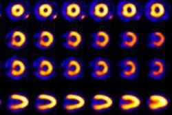
 |
| Images of a 50-year-old man acquired in six minutes at rest and four minutes of stress by Spectrum Dynamic’s D-SPECT. The images reveal a large ischemic abnormality involving the apex, anterior, septal, and apical inferior walls indicative of proximal left anterior descending stenosis. Images are courtesy of Spectrum Dynamics and Oregon Heart & Vascular Institute Sacred Heart Medical Center in Springfield, OR. |
A Stanford University study published in 2009 confirmed the capabilities of the SPECT technology to reduce imaging time for gated SPECT to as much as one-tenth the time of conventional SPECT.
"You do not compromise on image quality, because at the heart of [D-SPECT] are the solid-state detectors that are very efficient at capturing the counts coming out of the chest and a fundamental redesign from standard gamma-camera technology with multiple pivoting solid-state detectors that surround the chest," said Gambhir, who co-authored the 2009 study and helped develop D-SPECT. "This new design has proved to enhance spatial resolution, while still achieving a decrease in scan time."
A second SPECT system to offer a new system design was developed by GE Healthcare. The Discovery NM 530c features Alcyone technology, which consists of 19 pinhole collimators, each with four solid-state CZT pixilated detectors.
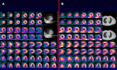 |
| Patient underwent rest-stress technetium-99m tetrofosmin myocardial perfusion imaging using 370 MBq for rest and 1,110 MBq for stress. Rest images appear below stress images. (A) On Discovery NM 530c SPECT system, rest and stress acquisitions were four and two minutes, respectively, and show fixed inferobasal wall. (B) With the addition of attenuation correction, increased tracer uniformity is seen throughout left ventricle, and inferior wall is seen to be normally perfused. Images courtesy of the Journal of Nuclear Medicine. |
Simultaneous imaging
The ability of the 19 collimators to image the heart simultaneously with no moving parts during data acquisition is designed to improve overall sensitivity, and give complete angular data for both dynamic studies and for the reduction of motion artifacts. "It gives you all sorts of advantages for flow, dynamics, and reducing artifacts, if the patient moves," Garcia said.
CardiArc is developing a cardiac SPECT device with three detectors that include a curved sodium iodide crystal and are set side-by-side to cover a 180° angle. The configuration is designed to help eliminate overlap in acquisitions from adjacent images, and it can reduce the time for a stress scan to as few as three minutes.
"We can reduce the time for a stress scan to approximately three minutes or can do the scan slower with less tracer," said Jack Juni, MD, CardiArc chairman and CTO and a physician at William Beaumont Hospital. "In initial tests, we have been able to reduce the radiation dose from the tracers by a factor of four."
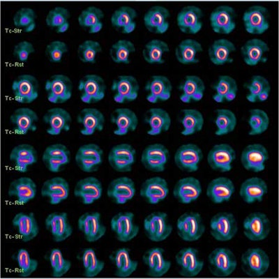
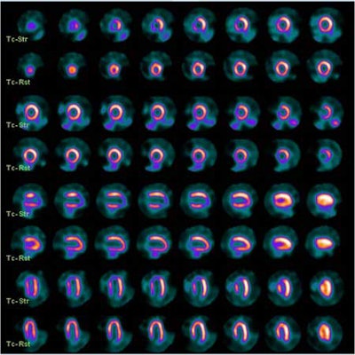
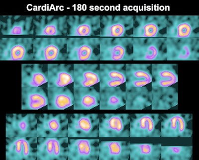 |
| Patient images taken with CardiArc HD-SPECT scanner. Images courtesy of CardiArc and Emory University School of Medicine. |
Small footprint
With a small footprint of 1 x 1.5 m, the CardiArc system is currently being validated at Emory University, with approximately 40 patients enrolled in the trial so far. Early results indicate that CardiArc detects the same lesions and the images "look at least as good and usually better than regular imaging," Juni said.
Also helping to transform cardiac SPECT, according to the study, is Digirad. The company was one of the first developers to use solid-state electronics and more than two detectors to simultaneously image the heart with its Cardius 3 XPO.
The system uses 768 pixilated, thallium-activated cesium iodide crystals coupled to individual silicon photodiodes and digital Anger electronics to create the planar projection images used for reconstruction.
Three detectors are fixed using a 67.5° angular separation, while patients are rotated through an arc of 202.5° sitting on a chair with their arms resting above the detectors. The typical acquisition time for a study is 7.5 minutes.
Enhanced sensitivity
Siemens Healthcare is offering its IQ-SPECT technology, which features two collimators mounted on the company's conventional dual-detector SPECT cameras, separated by 90°, and rotated around a patient to obtain a 180° reconstruction arc.
For increased sensitivity and resolution, the fields-of-view of these collimators are most convergent at their center, whereas the convergence is relaxed toward the edge of the field-of-view. The advantage of this approach is that it can be used by Siemens' existing dual-detector systems. Typical myocardial perfusion imaging acquisition times range from four to five minutes.
Philips Healthcare, meanwhile, offers BrightView XCT, a scalable SPECT/CT system designed for nuclear medicine and nuclear cardiology. The system combines BrightView SPECT and Philips' flat-detector x-ray CT for low patient dose levels and attenuation correction to reduce artifacts and exam times.
Future developments
Now that much of newer cardiac SPECT technology has been validated, Emory's Garcia said the next step is to develop appropriate imaging protocols to balance scan time and radiotracer dose. "It is becoming more patient-centric imaging," he added. "Instead of having one protocol in one lab, you probably have a few protocols that are geared to reducing radiation and improving images for the patient."
In recent years, the market has been rocky for cardiac SPECT, as uncertainty over reimbursement and the poor economy have led to less capital spending and delays in equipment purchases.
"This last year, with the combination of the economy and uncertainty about healthcare at the national level, sales of machines for everybody dropped to almost nothing," reflected CardiArc's Juni. "We are still in that [trend] right now. Physicians are trying to decide whether or not things are stable enough to invest."
Market rebound
Still, Juni is hopeful that the market might rebound in the next six to 12 months, if there is more word on reimbursement levels for the next three to five years. "Right now, reimbursement is only known with any certainty for 2011," he said. "For 2012, there were some scary numbers floated and no one really knows. That uncertainty is causing a lot of hesitation in the market."
If SPECT has a future in nuclear cardiology and there is a good return on investment, Garcia believes the technology can also be beneficial in imaging cancer and for other metabolic imaging applications. "We tend to think of molecular imaging more in terms of PET than SPECT, but there are many single photon agents that are very useful for molecular imaging," he added.
Stanford's Gambhir concurred, saying SPECT, like all of nuclear medicine, isn't all about the hardware; it's also about the tracer. "There are relatively good tracers for cardiac perfusion, like sestamibi and thallium," he said. "What's needed are better ways to do more than just perfusion assessment -- that is, viability assessment and, ideally, a 'one-stop shop' for SPECT where you can get both the coronary and cardiac anatomy, perfusion and viability, all in one shot."
| Authors of the Journal of Nuclear Medicine study received a research grant from GE Healthcare to evaluate the Discover NM 530c SPECT system. |


