Nuclear medicine firm UltraSPECT is touting study results that suggest its wide-beam reconstruction (WBR) technology reduces the required radiopharmaceutical dose and image acquisition time by 50% for SPECT myocardial perfusion imaging, when compared with conventional techniques.
The study was published in the March-April 2011 issue of the Journal of Nuclear Cardiology.
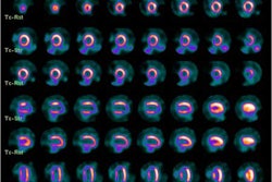
Researchers at St. Luke's-Roosevelt Hospital and Columbia University College of Physicians and Surgeons found that with either half the dose of technetium-99m sestamibi or half the acquisition time, WBR resulted in image quality superior to image processing with a technique based on ordered subset expectation maximization (OSEM).
The study evaluated images from 156 patients undergoing myocardial perfusion SPECT with a standard full-time acquisition protocol processed with routine OSEM methods. The same 156 patients underwent half-time acquisition, and the data were processed with the WBR algorithm.
A second study group of 160 patients received half of the standard radiopharmaceutical dose, with images acquired for the full standard acquisition time. These images were processed using WBR only.
Overall, WBR half-time and half-dose image quality was judged as superior to OSEM image quality in both segments of the study. There was no statistically significant difference between the two SPECT protocols in identifying the extent or severity of perfusion defects.


