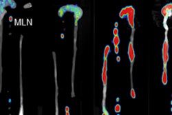
A group from the University of Münster in Germany has found that PET/CT may be able to more effectively detect and monitor inflammatory bowel disease such as Crohn's disease and ulcerative colitis, according to research published in the May issue of the Journal of Nuclear Medicine.
Typically, endoscopy and histology are used to identify lesions, but in this study PET/CT was able to identify lesions in mice along the complete intestinal wall that could be missed with traditional imaging techniques.
For the first phase of the study, lead study author Dr. Dominik Bettenworth and colleagues induced dextran sodium sulfate (DSS) colitis in mice and assessed FDG uptake in multiple sections of the colon.
Compared with control mice, an increase in FDG uptake was clearly noted in the DSS mice, with the most intense areas of uptake in the medial and distal colon. Significant correlation was found between the PET/CT and histologic results. The results also showed extraintestinal alterations, such as bone marrow activation, in the DSS colitis-induced mice.
Based on the results in the colitic mice, Bettenworth and colleagues identified the extent of mucosal damage as the parameter that correlated best to FDG uptake in the mice and in human patients with Crohn's disease. FDG-PET/CT scans of 25 Crohn's patients were retrospectively analyzed, and the findings were correlated toendoscopic procedures performed before or after CT with no treatments.
As with the DSS colitis results, increased FDG uptake was found in 87% of deep mucosal ulcerations, whereas mild endoscopic lesions were detected in approximately 50% of patients.


