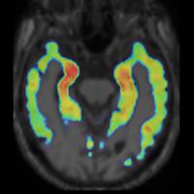
ST. LOUIS - A PET image using the novel tracer F-18 THK5117 to label tau protein and neurofibrillary tangles associated with Alzheimer's disease has received the Society of Nuclear Medicine and Molecular Imaging's (SNMMI) 2014 Image of the Year award.
The image from Japanese researchers at Tohoku University School of Medicine was selected from more than 1,700 studies presented during this year's annual meeting.
Dr. Nobuyuki Okamura and colleagues administered THK5117 to eight Alzheimer's patients and six age-matched healthy control subjects who underwent PET scans for 90 minutes.
PET scans were also performed with the amyloid PET tracer carbon-11 (C-11) Pittsburgh Compound B (PiB) on the same population to compare the selective binding ability of THK5117.
The researchers calculated standardized uptake value ratios (SUVRs) 60 to 80 minutes postinjection for THK5117 and 40 to 70 minutes postinjection for PiB using the cerebellar cortex as the reference region.
The Alzheimer's subjects showed THK5117 retention in the lateral and medial temporal cortices, which are known to contain high concentrations of tau deposits, according to the researchers. Patients are known to accumulate beta-amyloid plaque and tau proteins in the brain years before the onset of Alzheimer's disease.
THK5117 SUVR plateaued 60 minutes after injection, and the tracer's regional distribution differed considerably from that of PiB in patients with Alzheimer's.
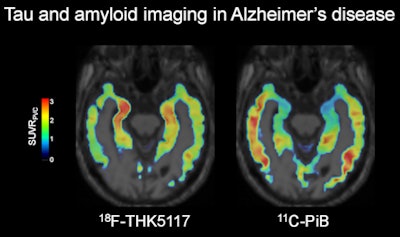 Image courtesy of SNMMI.
THK5117 retention in the temporal cortex correlated with the severity of dementia, and retention in the hippocampus (red area in the above image) correlated with hippocampal volume in Alzheimer's patients. THK5117 retention was also observed in the right temporal lobes of healthy control subjects with right temporal lobe atrophy.
Okamura and colleagues hope the THK5117-PET imaging technique will contribute to the development of new antidementia drugs, as the results provide information about the pathophysiology of Alzheimer's disease.


