With an eye toward lowering radiation exposure, a group of researchers found that an ultralow-dose high-efficiency SPECT scan for myocardial perfusion imaging can achieve better image quality than standard low-dose SPECT, according to a study published in the September Journal of Nuclear Medicine.
The researchers also concluded that assessment of extracardiac activity, overall perfusion, and ejection fraction was comparable between the scans, and this was accomplished at an ultralow dose of 1 mSv -- less than half the dose of standard low-dose SPECT.
Based on the results, lead author Dr. Andrew Einstein, PhD, an associate professor of medicine at Columbia University, and colleagues believe an ultralow-dose high-efficiency SPECT scan is feasible for stress myocardial perfusion imaging.
First of its kind
SPECT myocardial perfusion imaging plays an important role in the diagnosis of coronary artery disease, but concerns remain about the potentially adverse effects of radiation on cardiac patients, the authors noted.
"Compared with standard SPECT cameras, new high-efficiency cameras with specialized collimators and solid-state cadmium-zinc-telluride detectors offer potential to maintain image quality, while reducing administered activity and thus radiation dose to patients," wrote Einstein, along with colleagues from Brigham and Women's Hospital and several other institutions (JNM, September 2014, Vol. 55:9, pp. 1430-1437).
The researchers believe their study is the first to assess image quality, overall perfusion assessment, ejection fraction, and other factors in patients receiving ultralow-dose imaging using a high-efficiency SPECT camera, compared with standard low-dose SPECT imaging in the same patients.
A total of 101 patients with suspected or known coronary artery disease were prospectively enrolled at three sites for the study. All of the patients were scheduled to undergo SPECT myocardial perfusion imaging (MPI) for clinical indications. Subjects were excluded from the study if they were diagnosed with uncontrolled heart failure or hypertension or were obese.
To ensure a sufficient number of resting perfusion defects, the researchers created two groups. One included patients with no history of coronary artery disease and no known prior myocardial infarction or coronary revascularization. The second group included patients with a history of myocardial infarction, specifically hospitalization for acute myocardial infarction.
Each patient received two types of resting scans: an ultralow-dose high-efficiency SPECT study (D-SPECT, Spectrum Dynamics) and a standard low-dose SPECT scan (e.cam or Symbia, Siemens Healthcare; Forte, Philips Healthcare).
Ultralow-dose SPECT was performed with the patient resting in the supine position 45 minutes after administration of approximately 130 MBq (3.5 mCi) of technetium-99m (Tc-99m) sestamibi, with an effective radiation dose of 1.15 mSv. Image acquisition time was based on a patient's body mass index and ranged from 10 to 15 minutes.
Standard low-dose SPECT was performed 45 minutes later after approximately 278 MBq of additional Tc-99m sestamibi was injected, with an effective radiation dose of 2.39 mSv. Image acquisition time was 12 to 16 minutes.
"Single-injection rest imaging was chosen as the method with which to explore the low-radiation-dose procedure because it made it possible to perform ultralow-dose high-efficiency SPECT imaging under conditions identical to standard low-dose SPECT imaging, using a divided rest dose," the authors explained.
Two nuclear cardiologists assessed, among other variables, image quality, extracardiac activity, and overall perfusion of the two techniques.
'Excellent' results
The two readers rated image quality as "excellent" in 48 of the 101 ultralow-dose high-efficiency SPECT studies, compared with 24 of 101 for standard low-dose SPECT. Standard low-dose SPECT, however, had slightly more "good" images than ultralow-dose high-efficiency SPECT (48 versus 41, respectively). The results were similar among patients with and without suspected or known coronary issues.
| Image quality and extracardiac activity by SPECT type | ||||
| Excellent image quality | Good image quality | No extracardiac activity | Minimal extracardiac activity | |
| Standard-dose SPECT studies | 23.8% | 47.5% | 39.6% | 34.7% |
| Ultralow-dose SPECT studies | 47.5% | 40.6% | 54.4% | 26.7% |
Ultralow-dose high-efficiency SPECT detected no extracardiac activity in 55 patients, compared with 40 for standard low-dose SPECT. However, standard low-dose SPECT found more individuals with minimal or mild extracardiac activity (35 and 19, respectively) than ultralow-dose high-efficiency SPECT. The latter found 27 people with minimal extracardiac activity and 10 with mild extracardiac activity.
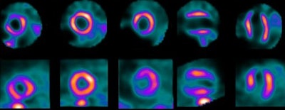
 Images of ultralow-dose high-efficiency SPECT (top row) and standard-dose SPECT (bottom row) at rest from a patient at Brigham and Women's Hospital, one of the three sites that participated in the study. Image courtesy of JNM.
The readers reached similar conclusions regarding overall perfusion assessment, with no significant difference between ultralow-dose high-efficiency SPECT and standard low-dose SPECT. Regarding ejection fraction, there was a statistically but not clinically significant difference between the two scan types, with strong correlation.
An ultralow-dose SPECT myocardial perfusion imaging study with a mean radiation dose of 1 mSv can achieve "high correlation" with conventional SPECT in terms of perfusion and function, with "improved image quality and comparable extracardiac activity," Einstein and colleagues concluded.
The results also suggest that a stress-only procedure is feasible with a high-efficiency SPECT camera and effective dose of 1 mSv, they added. Such an option would be "of interest to a broad community of physicians performing and ordering nuclear imaging studies."
Study disclosures
The study was supported by a grant from Spectrum Dynamics. Einstein has received grants for other research from GE Healthcare and Philips Healthcare. Two other co-authors are shareholders in Spectrum Dynamics and one other co-author is a consultant for Spectrum Dynamics.


