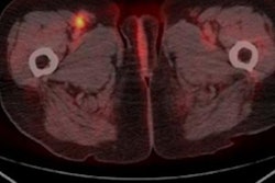
While the combination of PET and MRI has shown promise for detecting and characterizing some forms of cancer, the hybrid modality does not match the accuracy of invasive sentinel lymph node biopsy in melanoma patients, according to a study presented at ECR 2018 in Vienna.
Even with the addition of a diffusion-weighted MRI (DWI-MRI) sequence, PET/MRI offered only subpar sensitivity and positive predictive value in this clinical application, researchers in Germany found.
"The results are quite sobering," said lead author Dr. Benedikt Michael Schaarschmidt from the University of Düsseldorf. "We can surely state that PET/MRI cannot replace sentinel lymph node biopsy for N-staging in melanoma patients."
Frequent metastases
Lymph node metastases are quite common in patients undergoing treatment for melanoma, appearing in approximately 20% of all individuals with the disease.
"The latest guidelines demand sentinel lymph node biopsy as a method for N-staging," Schaarschmidt told ECR attendees. "But it is quite problematic because sentinel lymph node biopsy has a false-negative rate of close to 30% for all patients, and it is an invasive procedure. Therefore, we need a more reliable staging tool."
FDG-PET/CT has been used for various cancers; however, previous research has shown that it has very low sensitivity for lymph node metastases, especially in melanoma patients. Therefore, it has to be considered less reliable than sentinel lymph node biopsy.
In comparison, PET/MRI provides more detail than PET/CT, and it has high sensitivity for metastases. With MRI, clinicians can also include DWI in cancer-related protocols to enhance sensitivity with a high-resolution morphological image.
"So the question of the study was: Can integrated FDG-PET/MRI replace sentinel lymph node biopsy for lymph node staging in patients suffering from malignant melanoma?" Schaarschmidt said.
Patient analysis
The study included 52 patients: 30 women and 22 men with a mean age of 50.5 years. All of the patients underwent lymphoscintigraphy and SPECT/CT prior to sentinel lymph node biopsy. Of the 136 resected lymph nodes, 87 could be identified on SPECT/CT.
In all, 17 metastatic lymph nodes were detected by histopathology. Of those, four were smaller than 0.1 mm, seven were between 0.1 mm and 1 mm, and six were larger than 1 mm.
The researchers then correlated the sentinel lymph node results with those from PET/CT, PET/MRI, and PET/DWI-MRI.
Using the histopathological results as the reference standard, one radiologist and one nuclear medicine physician trained in hybrid imaging analyzed the imaging results. The pair rated the images on a five-point scale, with 1 designated as definitely benign and 5 as definitely malignant.
The PET/CT images were analyzed visually by looking at tracer uptake and quantitative standardized uptake values (SUVs), compared with the background uptake. The PET/MRI results were evaluated by tracer uptake and morphological features such as the shape of the lymph node, lymph node contour, and central necrosis, as well as DWI values in comparison to the background.
The researchers found that all three hybrid imaging modalities had poor sensitivity, positive predictive value (PPV), and negative predictive value (NPV). The addition of DWI to PET/MRI was not advantageous for sentinel lymph node metastases.
| Detection of lymph node metastases by modality | ||||
| Modality | Sensitivity | Specificity | PPV | NPV |
| PET/CT | 17.7% | 95.6% | 50.0% | 82.3% |
| PET/MRI | 23.5% | 96.9% | 66.7% | 82.3% |
| PET/MRI with DWI | 0% | 93.3% | 0% | 82.4% |
"For PET/CT, the results are just as bad as we would have expected them from previous literature, but PET/MRI did not do very well here," Schaarschmidt said. "The sensitivity is still very low and the positive predictive value and negative predictive value is too low to replace sentinel lymph node biopsy."
The poor performance of PET/MRI with DWI was due to contradictory information from PET and DWI, which reduced the number of true-positive findings. There also were two additional false positives with DWI, which further decreased the "already bad values of PET/MRI," he said.
There were no significant differences between benign and malignant lymph nodes based on tracer uptake, lymph node size, and morphology, according to the researchers.
"In conclusion, PET/MRI is not superior to PET/CT in lymph node metastases detection in patients suffering from malignant melanoma," Schaarschmidt concluded.


