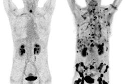
PET/MRI with the radiopharmaceutical gallium-68 (Ga-68) prostate-specific membrane antigen-11 (PSMA-11) is on par with currently available tools for determining the risk of prostate cancer progression, according to a study published in the December issue of the Journal of Nuclear Medicine.
The U.S. and German researchers also suggested that whole-body imaging with Ga-68 PSMA-11 PET/MRI could help physicians provide individualized treatment strategies for patients with prostate cancer.
In the retrospective study of 73 patients, risk for advanced prostate cancer was predicted using the Memorial Sloan Kettering Cancer Center (MSKCC) nomogram, the Partin tables, and Ga-68 PSMA-11 PET/MRI. Staging predictions were then compared with histopathologic results for three advanced disease states: lymph node metastases, extracapsular extension, and seminal vesicle invasion (JNM, December 2018, Vol. 59:12, pp. 1850-1856).
PET/MRI with Ga-68 PSMA-11 achieved an equivalent positivity rate for all three disease types, compared with the MSKCC nomogram and the Partin tables. Sensitivity and specificity were also comparable among the methodologies.
"Our results showed that PSMA-targeted PET/MRI performed equally well to established clinical nomograms for preoperative staging in high-risk prostate cancer patients and provided additional information on tumor location," said study co-author Dr. Andrei Gafita from the Technical University of Munich. "Translated into a clinical setting, the use of this imaging technique for preoperative staging might support treatment planning that may lead to improved patient outcomes."


