Blood oxygen level dependent (BOLD) MRI can provide an accurate, real-time way to monitor photodynamic therapy of solid tumors, according to investigators from The Weizmann Institute of Science in Rehovot, Israel.
Photodynamic therapy (PDT) is a relatively new approach to the treatment of tumors and nonmalignant hyperproliferative disease, such as opthalmic age-related macular degeneration, explained Dr. Shimon Gross and colleagues in a Nature Medicine article. PDT relies on the photoexcitation of an in vivo photosensitizing drug, at a wavelength matched to photosensitizer absorption, leading to tumor vascular occlusion, statis, necrosis and, ultimately, eradication.
However, "precise light delivery is essential for safe PDT...real-time imaging of the light impact and tissue response to PDT is presently unavailable...We therefore examined whether photoconsumption of oxygen and the consequent hemodynamic effects inherent to antivascular PDT could generate detectable BOLD contrast, thus enabling functional MRI of a photochemical process," the authors wrote (Nature Medicine, October 9, 2003, Vol. 9:10, pp. 1327-1331).
A solid tumor model was used for this study. PDT was paired with a photosynthetic pigment called palladium-bacteripheophorbide (TOOKAD, Steba Biotech, Toussus Le-Noble, France). The authors first assessed changes in tumor perfusion before, during and after TOOKAD-PDT with fluorescence intravital microscopy. They found that TOOKAD-PDT administration produced a rapid decrease in perfusion rate, reaching stasis in 3-4 minutes. In addition, spectral analysis indicated rapid photochemical oxygen consumption, leading to a decline in hemoglobin saturation levels.
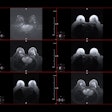
Gradient echo and MEGE images were obtained on a horizontal, 4.7-tesla spectrometer (BioSpec, Brucker BioSpin, Billerica, MA). BOLD-MRI was performed pre- and post--TOOKAD-PDT of a subcutaneous melanoma tumor.
"The strongest decrease in (T2-weighted) MR signal intensity (MRSI) was observed at the tumor rim...no detectable changes change in MRSI was observed in unilluminated muscle," they wrote. "These results suggest that rapid, local, photosensitized magnetic resonance BOLD contrast is generated during PDT."
The authors concluded that TOOKAD-PDT generated an appreciable attenuation (25%-40%) of the MR signal just at the targeted tumor site. In addition, observed changes in BOLD contrast is the result of photochemical and hemodynamic effects exclusive to the bloodstream, where photosensitization took place.
"Photosensitized BOLD MRI could provide accurate 3-D monitoring of tumor vascular insult, the hallmark of antivascular PDT...the concept of photosensitized functional MRI could be applied to guided-light delivery, which would permit interactive adjustment to the direction and intensity of the light beam," they stated.
By Shalmali PalAuntMinnie.com staff writer
November 10, 2003
Related Reading
Oxygen-influenced MRI helps detect viable myocardium, July 2, 2003
Laser ablation of liver metastases boosts long-term survival, March 7, 2003
New MRI method detects regional differences in myocardium, March 5, 2003
Copyright © 2003 AuntMinnie.com


.fFmgij6Hin.png?auto=compress%2Cformat&fit=crop&h=100&q=70&w=100)


.fFmgij6Hin.png?auto=compress%2Cformat&fit=crop&h=167&q=70&w=250)


