CHICAGO - Ultrasound gel can pull double-duty by enhancing MR images, according to a group from Brazil. The team found that using the gel could increase the confidence level in vaginal involvement when staging cervical cancer on pelvic MR.
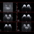
The researchers from the Federal University of Sao Paolo imaged 50 patients on a 1-tesla scanner (Magnetom, Siemens Medical Solutions, Erlangen, Germany). Half the patients were asked to self-administer 20 mL of standard ultrasound gel using a syringe. The other half underwent MRI without the gel. None of the women in the first group reported any pain or discomfort, the authors stated in their poster presentation Tuesday at RSNA.
The imaging protocol include T2-weighted TSE sequence and T1-weighted gradient-echo transverse sequences. Axial, coronal, and sagittal T2-weighted TSE images were acquired in the vagina. Sagittal and axial T2-weighted imaging was done for the bladder and rectum. Axial T1-weighted and T2-weighted images were obtained in the parametrium.
Two readers, who did not know which women used gel, interpreted the images. Local staging was done for the vagina, rectum, parametrium, and bladder. The images were scored on a five-point system with one indicating that the MR exam was difficult to evaluate and five representing an unequivocal evaluation.
The authors reported that using the intravaginal increased overall staging confidence, in addition to being simple and easily reproducible. In one case, the readers gave T2-weighted TSE sagittal and coronal images a rating of two for vaginal invasion. The same sequence with gel yielded a five score, indicating that there was no vaginal invasion.
Intraobserver confidence was analyzed using the Mann-Whitney U test. For the first reader, the Mann-Whitney U test was statistically significant for the vagina (p = 0.01), right and left parametrium (p = 0.03, for both), bladder (p = 0.007), and the rectum (p = 0.007). For the second reader, the results were also statistically significant, with p values ranging from 0.006 to 0.029 in the same areas.
"The most important advantage achieved was distension of the vagina and better delimitation of the tumor, especially on T2 sequences," the group wrote.
By Shalmali Pal
AuntMinnie.com staff writer
December 1, 2004
Related Reading
FDG-PET offers precise restaging info in recurrent cervical cancer, November 16, 2004
Demographics influence cervical cancer rates, July 28, 2004
Cervical cancer still cutting many lives short, April 22, 2004
Glucose metabolism regulators match FDG-PET uptake in cervical cancer, February 17, 2004
Copyright © 2004 AuntMinnie.com


.fFmgij6Hin.png?auto=compress%2Cformat&fit=crop&h=100&q=70&w=100)


.fFmgij6Hin.png?auto=compress%2Cformat&fit=crop&h=167&q=70&w=250)


