Women who have had breast conservation therapy have an increased risk of developing second breast cancers, and they require careful tracking. But which modality is most effective? MRI, according to a new study published in Radiology.
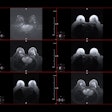
Annual mammography is recommended for women who have had breast conservation therapy, but other modalities have been explored because of mammography's sensitivity limitations and this group's higher-than-average risk of developing additional cancer. Researchers at Seoul National University College of Medicine in Korea found that using MRI to track these women detected 18 more cancers per 1,000 women than using mammography or ultrasound -- even in women with dense breast tissue.
Lead author Dr. Hye Mi Gweon and colleagues investigated single-screening breast MRI in women who had a history of breast conservation therapy and who had negative mammography and ultrasound findings six months before the MR exam. The study included 607 women who had 932 screening MR exams: 332 of the women had one round of MR screening, 225 had two rounds, and 50 had three rounds (Radiology, August 2014, Vol. 272:2, pp. 366-373).
At their institution, breast conservation therapy consists of breast-conserving surgery with radiation therapy, chemotherapy, hormonal therapy, or a combination of the three, depending on the patient, according to the authors. All 607 women had surgery plus radiation therapy, while 333 women also had chemotherapy and 387 also had hormonal therapy. After breast conservation therapy, the patients had bilateral mammography with ultrasound follow-up every six months for the first two years, with annual mammography and ultrasound thereafter.
All screening MR exams were done on a 1.5-tesla unit (Signa, GE Healthcare) with a dedicated breast coil.
Of the 607 first-round MR exams:
- 293 (48.3%) were BI-RADS 1
- 197 (32.5%) were BI-RADS 2
- 94 (15.5%) were BI-RADS 3
- 23 (3.8%) were BI-RADS 4
Of the BI-RADS 4 lesions, seven were biopsied under MR guidance and 16 under ultrasound guidance; the biopsies revealed 10 cancers and 13 benign lesions. Of the BI-RADS 3 lesions, one cancer was identified, for a total of 11 cancers found.
Gweon's group determined that the overall cancer detection rate of first-round MR exams was 18.1 per 1,000 women. Of the detected cancers, 72.7% were invasive ductal carcinoma and 27.3% were ductal carcinoma in situ.
Two cancers were detected in the second-round MR exams, and no cancers were found in the third-round MR exams. Finally, one false-negative was found at six-month follow-up mammography after the initial MR screening exam had identified a BI-RADS 2 lesion.
MRI's specificity was 82.2% and its sensitivity was 91.7% -- particularly gratifying results, as 80.2% of the women in the study had BI-RADS category 3 or 4 dense breast tissue, according to the group.
"This cancer detection rate is comparable to that of high-risk women, such as BRCA carriers or those with a lifetime breast cancer risk of 20% to 25% or greater," Gweon and colleagues wrote.
And the women were younger as well: Cancers were more frequently identified by MRI in women younger than 50 than in those older than 50, according to the researchers.
What's the bottom line? Using MRI to track women who have undergone breast conservation therapy finds more breast cancers, which is good news for women at risk for second cancers, and for those in this subgroup who have dense breast tissue, they concluded.


.fFmgij6Hin.png?auto=compress%2Cformat&fit=crop&h=100&q=70&w=100)


.fFmgij6Hin.png?auto=compress%2Cformat&fit=crop&h=167&q=70&w=250)


