Wednesday, November 30 | 12:15 p.m.-12:45 p.m. | W5A-SPGI-4 | Learning Center - GI DPS
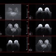
At this Wednesday afternoon poster session, researchers will present study results that highlight the benefits of using artificial intelligence with a contrast-enhanced T1-weighted MRI sequence for imaging the liver.Dr. Francesca Castagnoli of the U.K.'s Royal Marsden NHS Foundation Trust and colleagues assessed the effect of applying a neural network trained on high-resolution MRI images to a T1-weighted MRI sequence for the liver, comparing it with taking the same sequence without AI. Fifty patients underwent a contrast-enhanced liver MRI with gadolinium ethoxybenzyl-diethylenetriaminepentaacetic acid (Gd-EOB-DTPA); they were scanned with a conventional 17-second T1-weighted sequence; a 17-second T1-weighted sequence with the neural network assist; and with an accelerated, 12-second T1-weighted sequence with the neural network reconstruction. Two radiologists evaluated the overall image quality, contrast-to-noise ratio, lesion and vessel edge sharpness, and breathing motion artifacts.
The neural network improved scores for image quality, contrast-to-noise ratio, and vessel and lesion edge sharpness for the 17-second and 12-second acquisitions compared with standard image reconstruction -- and there was no statistically significant difference between the two acquisition lengths, the investigators found. Additionally, there was no difference between the number of lesions and the diameter of the smallest lesion identified using the 12-second neural network-enhanced sequence compared with the conventional 17-second acquisition.
As for respiratory motion artifacts, there were no differences between all three image sequences, the group noted.
"AI-augmented T1-weighted imaging can improve image quality [and] reduce image acquisition time without compromising diagnostic performance," the team concluded.


.fFmgij6Hin.png?auto=compress%2Cformat&fit=crop&h=100&q=70&w=100)

.fFmgij6Hin.png?auto=compress%2Cformat&fit=crop&h=167&q=70&w=250)


