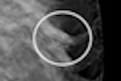
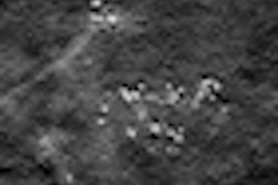
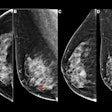
Digital breast tomosynthesis (DBT) appears comparable to clinical mammographic spot views for breast mass characterization, according to a new study published online in Radiology. If this finding is confirmed by future studies, using DBT in clinical practice could reduce the number of patient recalls and women's exposure to additional radiation.
Researchers at the University of Michigan compared DBT images and mammographic spot views of 67 masses -- 30 malignant and 37 benign -- and found that mass characterization in terms of visibility ratings, reader performance, and BI-RADS assessment with DBT was similar to that of spot views (Radiology, October 13, 2011).
Mammographic spot views, with or without magnification, have been a key tool for diagnostic breast imaging to identify masses, reduce noise from scattered radiation, reduce superimposition of overlapping tissue, and also improve the effective spatial resolution of the detector, which enhances tissue contrast, according to Dr. Mitra Noroozian and colleagues.
But with the advent of digital mammography and breast tomosynthesis, it has been suggested that tomosynthesis might improve mammographic sensitivity in similar ways. Such a development would benefit women by requiring fewer patient recalls and less radiation from follow-up exams. Therefore, Noroozian's team sought to compare the two modalities' ability to characterize breast masses as benign or malignant.
Four radiologists blinded to whether images were spot views or DBT individually evaluated the images in random order. The readers rated the images for visibility (10-point scale), likelihood of malignancy (12-point scale), and BI-RADS classification. Masses categorized as BI-RADS 4 or 5 were compared with histopathology data to determine true-positive results for each modality.
DBT images were acquired with a combined DBT and whole-breast research system (GE Global Research). The spot views were defined as a clinically obtained spot compression or spot magnification views, acquired either with digital or analog units (analog images were digitized), and if multiple spot views in the same projection were available to be matched to a DBT image, the single best view was chosen. From the 67 masses, Noroozian's team gathered 93 matched image sets for review. DBT images were cropped to display a volume of tissue that approximated the tissue visible on the matching spot view.
All four study readers reported slightly better mean mass visibility with DBT images compared with spot views, although only one reader's results achieved statistical significance (DBT range, 3.2 to 4.4; spot view range, 3.8 to 4.8), Noroozian and colleagues wrote. The area under the receiver operator characteristics (ROC) curve ranged from 0.89 to 0.93 for DBT and 0.88 to 0.93 for spot views, while the partial area index above a sensitivity threshold of 0.90 ranged from 0.36 to 0.52 for DBT and 0.25 to 0.40 for spot views.
The readers characterized seven additional malignant masses as BI-RADS 4 or 5 with DBT than with spot views, at a cost of five false-positive biopsy recommendations, with a mean of 1.8 true-positive and 1.3 false-positive assessments per reader, the authors wrote.
"Our results imply that when DBT becomes integrated into clinical breast imaging practice, [spot views] might not be necessary for mass assessment," they wrote. "Vendors of clinical DBT prototypes have achieved or will achieve radiation doses similar to that of screening mammography. ... If future studies confirm that DBT can be integrated into screening protocols and that DBT can substitute for [spot views] to characterize masses, then women may be spared from being recalled for diagnostic views and from the associated incremental radiation exposure."
Noroozian's group conceded that their study had limitations, including a small number of masses and the mix of spot view images, which resulted in a nonuniform comparison between spot views and DBT.
"DBT may or may not obviate [spot views] in the diagnostic workup of a breast mass," they wrote. "Larger studies with more diverse mammographic findings and full sets of diagnostic mammographic images may better define the emerging role that DBT might play in clinical breast imaging practice."


