Tuesday, December 2 | 3:30 p.m.-3:40 p.m. | SSJ01-04 | Arie Crown Theater
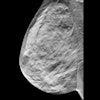
Researchers have found that a dual-energy breast imaging technique may be able to decrease the number of unnecessary breast biopsies.Three-compartment breast imaging is a dual-energy mammography technique that utilizes standard mammography equipment and an in-image phantom that mimics different thicknesses of the three tissue components (water, lipid, and protein), explained presenter Karen Drukker, PhD, of the University of Chicago. At the cost of only a 10% increase in radiation dose over a standard mammogram, this method enables the calculation of tissue composition throughout the breast.
The group set out to investigate whether it's possible to move beyond x-ray mammography and obtain additional information from the tissue composition throughout the breast and especially within a lesion and its periphery, Drukker said. Even though screening mammography has a track record of saving lives, some studies have found that 60% of women receiving these studies will have a false-positive result.
"This is where we believe three-compartment imaging will help," she said. "It is intended as a diagnostic method, after detection of a mammographically suspicious finding, to avoid unnecessary biopsies and increase the positive predictive value of biopsy."
The group's preliminary study found that there are specific tissue composition "signatures" for different lesion types.
"Invasive cancers, for example, tend to have high water content within and also in the immediate surrounding tissue," she told AuntMinnie.com.
The measurements obtained from the three-compartment images were largely independent from those obtained from the regular mammograms. Combining information from both modalities significantly improved the ability to distinguish between benign and malignant lesions, Drukker said.
"Hence, to date ... results are promising that three-compartment breast imaging can live up to the expectations of helping to avoid unnecessary biopsies in healthy women, while ensuring that women who do have breast cancer get timely and appropriate follow-up," Drukker said.


