Dynamic contrast-enhanced MRI (DCE-MRI) can help distinguish women with high recurrence risk of breast cancer from those with low recurrence risk, a study published January 23 in Radiology found.
Researchers led by Dooman Arefan, PhD, from the University of Pittsburgh found that quantitative background parenchymal enhancement (BPE) found on DCE-MRI combined with tumor radiomics could be an alternative to the Oncotype DX recurrence score in assessing breast cancer recurrence risk.
“This finding has particular clinical value considering that the Oncotype DX recurrence score is expensive and unavailable in many low-resource health care settings,” Arefan and colleagues wrote.
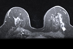
BPE refers to the contrast enhancement of normal breast tissue on DCE-MRI and is known as a risk factor for breast cancer development. However, the researchers noted that it remains unexplored how quantitative BPE measures are tied to outcomes for women receiving breast cancer treatment.
In women with early-stage, estrogen receptor-positive, node-negative invasive breast cancer, oncologists use the Oncotype DX recurrence score (Genomic Health) to analyze the expression of 21 genes to predict the risk of distant recurrence. The resulting recurrence score helps oncologists decide on appropriate treatment plans.
Arefan and co-authors investigated whether quantitative BPE measurements in one or both breasts could be used to predict recurrence risk in breast cancer patients. They used the Oncotype DX recurrence score as the reference standard.
The team included data collected between 2007 and 2012 from 187 women with breast cancer. Of the total, 127 were included in a development set (33 with high risk and 94 with low- or intermediate risk). These women had an actual local or distant recurrence rate of 15.7% at a minimum 10 years of follow-up. The team also included 60 women in a test set (16 with high risk and 44 with low- or intermediate risk).
The researchers found significant associations between BPE measurements quantified in both breasts and increased odds of a high-risk Oncotype DX recurrence score. This included an odds ratio range of 1.27 to 1.66 (p<0.001 to p = 0.04).
Additionally, BPE measures on DCE-MRI combined with tumor radiomics helped determine which patients had a high-risk Oncotype DX recurrence score versus those with a low- or intermediate-risk score. This included an area under the curve (AUC) of 0.94 in the development set and 0.79 in the test set. This combined approach also achieved high negative predictive values of 0.97 and 0.93 in predicting actual distant recurrence and local recurrence, respectively.
The study authors called for future studies on larger multi-institutional patient cohorts to validate their results.
In an accompanying editorial, Wenhui Zhou, MD, PhD, from Stanford University and Habib Rahbar, MD, from the University of Washington in Seattle wrote that the Arefan team’s results “lay the groundwork and present the exciting prospect for developing a robust and high-performing imaging method that is capable of predicting breast cancer recurrence within diverse real-world settings across different clinical practices.”
“In the current era of precision oncology, a data-driven platform to aggregate and synthesize imaging biomarkers holds tremendous potential in personalizing risk stratification and guiding clinical decisions,” they added.
The full study can be found here.


