Minnies winners, page 3
Best New Radiology Device
Winner: Aquilion One Vision 640-slice CT scanner, Toshiba America Medical Systems
In a year in which CT swept most Minnies award categories, it's only fitting that the prize for Best New Radiology Device should go to Toshiba America Medical Systems for its Aquilion One Vision scanner.
 Toshiba's Aquilion One Vision CT scanner. Image courtesy of Toshiba.
The new system, which debuted at the 2012 RSNA meeting, is the second generation of the groundbreaking Aquilion One 320-detector-row scanner first introduced in 2007. Toshiba's goal for Aquilion One Vision was to offer a CT scanner that enables the platform's wide 16 cm of anatomical coverage to be used in a broader range of environments, such as for obese patients or those with high heart rates presenting to the emergency room.
Aquilion One Vision sports a rotation speed of 275 msec; a wider gantry aperture of 78 cm, compared with 72 cm on the older model; and table support for patients up to 661 lb. The system uses Toshiba's ConeXact technology, which recontructs data in three dimensions, to acquire two sets of data at 0.5-mm slice widths with each gantry rotation.
Toshiba addresses the CT radiation dose question by making its adaptive iterative dose reduction (AIDR) 3D protocol available on the system, which received FDA clearance in 2012.
Researchers are already finding new uses for the scanner. For example, a study published in Radiology in January from the U.S. National Institutes of Health found that the scanner sharply reduced radiation dose while maintaining image quality. Median radiation dose was less than 1 mSv for more than 100 patients scanned with Aquilion One Vision, compared with 2.67 mSv using a first-generation 320-row system.
Runner-up: 3D breast biopsy option for Affirm device, Hologic
Best New Radiology Software
Winner: DoseTrack radiation dose monitoring software, Sectra
One of the most surprising revelations brought to light in the past several years in the radiation dose debate is that radiology professionals never really had an easy-to-use tool to notify them if a patient received too much radiation. That's starting to change as software developers create new applications to make radiation dose management much easier.
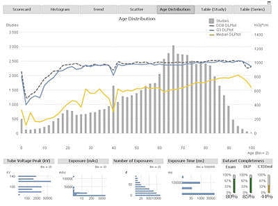
The winner of this year's Best New Radiology Software award, Sectra DoseTrack, demonstrates how much things are changing. The software enables imaging facilities to track and manage radiation dose across their enterprise, while also providing alerts if a patient receives too much dose from individual exams.
','dvPres', 'clsTopBtn', 'true' );" >
Click image to enlarge.
Sectra's DoseTrack software monitors radiation dose from all connected modalities. Image courtesy of Sectra.
Sectra DoseTrack allows practitioners to monitor radiation dose at the modality, exam, and patient level. The software can be configured to support local and national dose reference levels (DRLs) to help healthcare enterprises confirm that they're performing to expected thresholds. Data can be exported for further analysis in Excel or for reporting to regulatory entities such as the American College of Radiology's Dose Index Registry.
Specific imaging systems can be monitored to determine whether they are exposing patients to too much radiation, and the application also supports the follow-up of individual patients if a radiation incident is suspected. Other features include on-demand access to recent dose data, automated email reports and alerts, visualization of dose trends via dynamic graphs, and HL7 integration.
Sectra developed DoseTrack with Region Skåne, a 10-hospital network in southern Sweden, where it has been in use since 2008 to track dose on a regional scale. The company debuted the software as a commercial product at the 2012 RSNA meeting, and it registered DoseTrack with the FDA in July 2013 as a class I medical device.
Runner-up: ACR Select, National Decision Support Company
Best New Radiology Vendor
Winner: National Decision Support Company
While its ACR Select software came in second in the race for Best New Radiology Software, National Decision Support Company (NDSC) scored a nice consolation prize, being named the Best New Radiology Vendor by the Minnies expert panel.
NDSC was founded by an executive team that includes Michael Mardini, who has an impressive track record in founding imaging informatics companies. These include Talk Technology, which was bought by Agfa HealthCare in 2001, and Commissure, which was the recipient of the Minnies award for Best New Radiology Vendor in 2006 and was subsequently acquired by Nuance in 2007.
Mardini and the rest of his team are hoping lightning strikes again with NDSC, which is the exclusive distributor of ACR Select, a decision-support software platform based on the American College of Radiology (ACR) Appropriateness Criteria. The software attacks the problem of overutilization of advanced imaging exams, which is costing the healthcare system millions in unnecessary costs and, in the case of some modalities such as CT, exposing patients to unnecessary radiation.
The idea is that ACR Select puts the information needed for more appropriate ordering at the fingertips of referring physicians -- potentially replacing other utilization management tools such as radiology benefits managers.
','dvPres', 'clsTopBtn', 'true' );" >
Click image to enlarge.
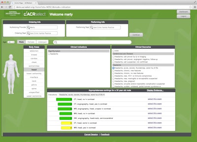
The ACR Select decision-support platform is based on ACR Appropriateness Criteria. Image courtesy of NDSC.
ACR Select went into commercial release in mid-2013, and NDSC has been focusing its attention on performing integrations with major electronic health record (EHR) platforms used by clinicians to order studies. Integrations have already been completed with software offered by Epic Systems, Cerner, and Allscripts, and they are underway with applications marketed by Meditech and Siemens Healthcare.
Referring physicians encounter ACR Select through their EHR software whenever they try to order an imaging exam. After entering data on the patient's condition, clinicians are presented with a structured list of indications that highlight the appropriate imaging studies for that condition.
Should a referring physician wish for additional consultation, NDSC has a module that enables a healthcare organization to connect the clinician with a radiologist, according to Bob Cooke, vice president of marketing and strategy at the company. ACR Select can also be used in an offline mode, apart from the exam-ordering process, as a reference tool for physicians to determine the appropriateness of a scan they are considering. Doctors can even use ACR Select to consult with patients on exams patients might want but which aren't clinically appropriate.
The latter features highlight how NDSC views ACR Select as more than simply a tool to nudge referring physicians to order less imaging.
"This is an incredibly powerful enabling tool to showcase how imaging can help improve patient care, as opposed to something focused on simply utilization management," Cooke told AuntMinnie.com. "We want to demonstrate that by using content we can drive imaging to a more appropriate level, and help radiologists define their role in the care cycle."
Runner-up: Montage Healthcare Solutions
Best Radiology Mobile App
Winner: OsiriX HD (iOS)
OsiriX HD traces its lineage to OsiriX, a popular, longtime open-source image viewer for the Mac. Since its debut in 2011 as an iPhone and iPad app, OsiriX HD has won many devoted fans, and it's a fitting first choice for the inaugural Best Radiology Mobile App award in the Minnies.
Developed by Swiss firm Pixmeo, OsiriX HD is a DICOM image viewer for iOS devices, including the iPhone, iPod touch, and iPad. It allows basic image manipulation such as zoom, pan, and window/level using gestures, and it supports standard DICOM communications such as DICOM Query and Retrieve. It also offers support for JPEG 2000 and JPEG DICOM image formats.
Version 3.6.2 of the app is available for $29.99; users must have at least iOS 4.3.
Runner-up: Radiology Assistant (iOS)
Best Radiology Image
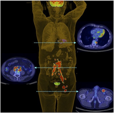
Winner: 3D PET digital reconstruction of Merkel cell carcinoma patient
A new category in the Minnies this year, the Best Radiology Image award goes to a PET scan of a patient with Merkel cell carcinoma, an aggressive skin disease that often reoccurs and carries a high risk of both nodal and distant metastases.
Researchers from Peter MacCallum Cancer Centre and the University of Melbourne acquired the image as part of a clinical study on the effectiveness of F-18 FDG PET in patients with Merkel cell carcinoma. They wanted to see how well PET could be used to stratify patients with the disease, which, while rare, nearly tripled in incidence in the U.S. between 1986 and 2001. Their results were published in the August 2013 issue of the Journal of Nuclear Medicine.
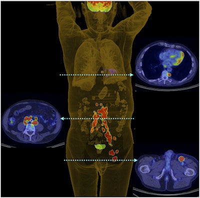 The winning image: 3D PET shows metastases in patient with Merkel cell carcinoma. Image courtesy of JNM.
Surgical resection is typically the most common treatment for Merkel cell carcinoma, but it has a high rate of locoregional failure if not followed by postoperative radiation therapy. PET has been used in small studies with promising results in improving patient management, so the group wanted to investigate in a larger population whether the modality could stratify patients and predict their prognosis.
In the study, 102 patients were staged using PET, and the researchers examined whether the scans resulted in a change in patient management. They found that the imaging technique did, in fact, change treatment for 38 patients, and PET altered staging for 22 patients.
The scan named as Best Radiology Image is a 3D digital PET reconstruction that shows left inguinal nodal and extensive retroperitoneal and thoracic nodal metastases in a patient with left-lower-limb Merkel cell carcinoma.
Runner-up: Wide-area coronary CT angiography with 640-slice reconstruction
Previous page | 1 | 2 | 3
