Dear Advanced Visualization Insider,
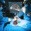
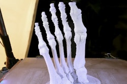
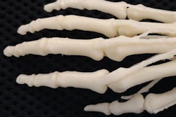
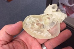
3D printing is growing by leaps and bounds, rapidly expanding its range of applications in surgical planning, training and education, and -- in the not-too-distant future -- even tissue and organ replacement, experts say. If the burgeoning multidisciplinary field has a problem, it may be in the "siloing" of different kinds of specialists in technology and healthcare, each working a piece of the industry with little feedback or input from experts in other fields.
But there won't be any silos on the horizon at next month's 3DHeals meeting in San Francisco. A broad array of clinical care professionals, industry leaders, and technology experts will gather at the April 20 meeting to share what they know about 3D printing: what's possible, what's being tried, and where things are heading in the growing field. Find out more by clicking here.
One group that's stretching the limits of what's possible in 3D printing is from George Washington University in Washington, DC. A team there used CT and MR images of the cerebellopontine angle and skull base to build a complex 3D-printed model that offers one of the first good examples of cranial nerves. Learn more about how they did it in this issue's Insider Exclusive, brought to you first as an Insider subscriber.
Farther afield, a group in Spain used deep-learning techniques, specifically convolutional neural networks, to build a computer-aided detection (CAD) system that offers first reads of chest x-rays in a fraction of the time it takes human readers to look over them, and with high accuracy for detecting abnormal results that need priority reading by humans. For more on this time-saver, click here.
In the Netherlands, investigators are using color mapping of the cerebral vasculature in 4D CT angiography to speed interpretation in the highly time-sensitive acute stroke setting, while potentially improving accuracy as well. Get the news from Nijmegen here.
Finally, a lung nodule study found that CAD handily beat a team of human readers in the detection of suspicious part-solid and nonsolid lung nodules. Find out how they did it here.
Along with nodules, long-term smokers often develop chronic obstructive pulmonary disease, which can now be quantified automatically.
And there's more -- right here in your Advanced Visualization Community. For the rest of the news in advanced visualization, 3D, and CAD, we invite you to scroll through the links below.

