CHICAGO -- Interstitial lung disease (ILD) involves more than just CT features, and it is time to start pushing boundaries with functional imaging, according to research presented December 2 at RSNA 2025.
One example of ILD research is using phase-resolved functional lung (PREFUL) MRI, a new technique that analyzes air movement in and out of the lungs and blood flow through the lungs, which doesn't require radiation or intravenous contrast. The technique may have potential to become a noninvasive diagnostic and monitoring tool, complementary to CT, for fibrosing ILD (F-ILD) in particular.
ILD involves diffuse lung disease characterized by pulmonary and interstitial fibrosis and the loss of alveolar capillary function, leading to gas exchange disorders in the lungs. Much is still unknown about ILD, why it develops, and how to stop the progression of lung fibrosis.
Ventilation (V) and perfusion (Q) are key to healthy functioning gas exchange. Yifei Ni from the Chinese Academy of Medical Sciences and Peking Union Medical College is exploring PREFUL MRI in V/Q dynamics in hopes of improving prognosis for those facing F-ILD.
For her session, Ni presented results from her PREFUL MRI research study that paired 30 F-ILD patients (16 with idiopathic pulmonary fibrosis [IPF] and 14 with another type of F-ILD; mean age, 64.6; 19 of the participants were male) with 30 healthy controls.
Ni and colleagues looked not only for correlation between PREFUL MRI-derived metrics and lung lesions on high-resolution CT but also for correlation of PREFUL MRI-derived metrics and pulmonary function tests in F-ILD patients.
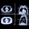
 Study results presented at RSNA 2025, highlighting phase-resolved functional lung (PREFUL) MRI-derived metrics.Liz Carey
Overall, PREFUL MRI demonstrated strong correlations with pulmonary function parameters, Ni reported.
The group also found correlations between PREFUL MRI-derived metrics and lung lesions on HRCT. Ni reported that mean perfusion is correlated with whole-lung percentage of honeycombing (r = 0.447, p = 0.013), reticulation (r = 0.404, p = 0.027), fibrotic lesions (r = 0.481, p = 0.007), and interstitial lung abnormalities (r = 0.601, p < 0.0001).
In addition, Ni reported that [perfusion defects in percent] QDP exclusive correlated with whole-lung percentage of honeycombing (r = 0.461, p = 0.01), reticulation (r = 0.41, p = 0.024), fibrotic lesions (r = 0.446, p = 0.013), interstitial lung abnormalities (r = 0.594, p < 0.001), and emphysema (r = 0.379, p = 0.039).
A number of papers have been published about PREFUL MRI, but the technique is currently not used for diffuse lung disease in the clinical setting, stated session moderator Michael McInnis, MD, a chest radiologist at the University of Toronto and Ni. However, Ni's research would be worth exploring in a clinical setting, McInnis added.
Also noted, this study of PREFUL MRI was not correlated with traditional V/Q scans using lung scintigraphy.
This session also highlighted the work of postdoctoral research fellow Tician Schnitzler, MD, and researchers from the University of California, San Francisco (UCSF) and Cantonal Hospital Aarau in Switzerland.
Schnitzler presented results from a multicontinental study of imaging biomarkers and clinical features of interstitial lung abnormalities (ILA) and their potential to predict progression to clinical IPF.
Supported by a 2024 RSNA research fellowship grant, the Swiss Society of Radiology, and the Gottfried and Julia Bangerter-Rhyner Foundation, the retrospective, multicenter study included 743 patients with CT-detected ILA (523 low-risk cases and 220 high-risk cases). Most ILAs are asymptomatic and do not progress to symptomatic ILD, but some ILAs do evolve into IPF, Schnitzler noted.
"Accurate risk stratification of ILA is critical for guiding follow-up and early intervention," Schnitzler said. First, the session highlighted that defining ILA is an important step. The study defined ILA as the following:
- Reticulation >5% of lung parenchyma (visually or quantitatively)
- Not severe enough to be ILD by imaging (no proximal traction bronchiectasis or honeycombing)
- Incidental (no definite clinical suspicion of symptomatic ILD)
- Subpleural component of reticulation (i.e., excludes fibrotic sequels of infection, for example)
In their study, Schnitzler and colleagues examined imaging features, including subpleural fibrotic reticulation, cranial extent of fibrosis, moderate and severe emphysema, anterior lung involvement, and central extent of fibrosis. The study reconfirmed that subpleural fibrotic reticulation is a strong predictor of IPF progression.
However, multivariate regression analysis excluded all features but the central extent of fibrosis and anterior lung involvement, which remained statistically significant as high-risk features, according to Schnitzler.
 Tician Schnitzler, MD, from the University of California, San Francisco (UCSF) and Cantonal Hospital Aarau in Switzerland presented results of a study of interstitial lung abnormality (ILA) imaging features predictive of disease progression.Liz Carey
The overall risk stratification model demonstrated "excellent discrimination" with an area under the curve (AUC) of 0.915, indicating strong predictive capability for identifying patients at high risk of ILA progression, Schnitzler reported, adding that the study included four external validation sites. Three in the U.S. included UCSF and Duke University, and Taiwan and Switzerland outside the U.S. AUCs were similar, Schnitzler said.
Although American Thoracic Society (ATS) guidelines recently have addressed ILA follow-up, such follow-ups should be an individual decision rather than dictated by guidelines, Schnitzler noted during the Q&A. However, Schnitzler emphasized ILA is an imaging finding (rather than a clinical diagnosis) but recommended follow-up with the presence of high-risk features.
McInnis remarked that ILAs are a controversial topic. Session moderator Dr. Mini Vithal Pakkal, also from the University of Toronto, said that the research findings will hopefully shed more light on how best to monitor these patients.
"One of the key messages was with ILA -- we need to look at additional parameters which will help us identify which patients are going to progress ... maybe some central fibrosis and anterior fibrosis we need to keep in mind," Pakkal said.
"I think the overall loud message for us was that ILD has more to it than just CT features, and we need to start pushing the boundary with functional imaging whether it is the PREFUL magnetic resonance imaging, whether it is deformation markers, whether it is looking for pulmonary vascular remodeling," Pakkal concluded.
For full coverage of RSNA 2025, visit our RADCast.