Technetium-MDP Chemistry & Pharmacology
Bone is a composite material of inorganic crystals bound to protein
[4]. The mineral phase, built of crystals containing mainly calcium and
phosphate, is called hydroxyapatite and this is bound to a matrix
largely consisting of a single protein- collagen [4]. Collagen provides
toughness to bone, making it less brittle so that is better resists
mechanical stress [4]. Healthy adult bone is in homeostasis- with a
constant rate of remodeling due to the activity of osteoblasts laying
down new bone and osteoclasts resorbing old bone in response to
mechanical stresses [4]. The bone consists of two components- a compact
(cortical) component and a cancellous (trabecullar) component that
serve different functions [4]. Although cortical bone forms most of the
bone mass (80%), it represents a minority of the bone surface which is
mostly trabecullar bone) [4].
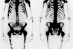
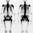
Technetium-MDP (Methylene diphosphonate) is the agent used for bone
imaging (a typical preparation of the radiopharmaceutical is 95% bound
to technetium). It is an
organic analog of pyrophosphate and contains an organic P-C-P bond.
Tc-MDP undergoes protein binding in the blood- only unbound tracer is
available for bone uptake [4]. The agent affixes to
the bone surface by the process of chemisorption- attaching to
hydroxyapatite
crystals in bone and calcium crystals in mitochondria. After
administration, it is
postulated that Tc-MDP dissociates into its technetium and MDP moieties
which are then
adsorbed onto the organic and inorganic (hydroxyapatite) phases,
respectively [2]. About 40-50% of the compound will be affixed to bone
by 3 to 4 hours after
injection. The adsorption of Tc-MDP to hydroxyapatite appears to
increase at low Ph [4].
Excretion is primarily renal [4] and 70% of the administered dose is eliminated by 6 hours. For an adult patient, the usual dose is 20 to 25 mCi. The whole body dose is 0.01 rad/mCi; the urinary bladder is the critical organ, receiving about 0.1-0.2 rad/mCi. The radiation dose from a Tc-MDP bone scan is approximately 4.2-5.3 mSv (420 mrem) [3,4].
The mechanism for increased uptake (increased activity) of the tracer in osseous lesions is multifactorial:
- Increased blood flow: Bone in hyperemic regions is exposed to more tracer over any given time period. Because the extraction of the radiotracer is non-linear and tends to plateau, increased flow will only result in an increase in bone activity compared to normal bone of about 1.5 to 2 times.
- Areas and rate of new bone formation (surface area): In these areas there is increased osteoid formation and increased mineralization of osteoid. Newly forming hydroxyapatite crystals are of smaller size than mature crystals and provide a greater surface area for binding.
- Interruption of sympathetic supply: See Reflex Sympathetic Dystrophy discussion.
REFERENCES:
(1) Radiographics 2003; Love C, et al. Radionuclide bone imaging: an illustrative review. 23: 341-358
(2) J Nucl Med 1993; Jan.: p.104
(3) J Nucl Med 2009; Iagaru A, et al. Novel strategy for a cocktail 18F-fluoride
and 18F-FDG PET/CT scan for evaluation of malignancy:
results of the pilot phase study. 50: 501-505
(4) J Nucl Med 2013; Wong KK, Piert M. Dynamic bone imaging with 99mTc-labeled
diphosphonates and 18F-NaF: mechanisms and applications.
54: 590-599
