Pulmonary Varix:
Clinical:
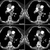
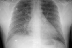
A non-obstructive dilatation of one or more pulmonary veins usually seen near the point of entry to the left atrium. The lesion is associated with pulmonary venous hypertension and left atrial enlargement (mitral valve disease most commonly, but also aortic coarctation, cirrhosis, or emphsema [6]), but it may also be congenital. With regards to mitral regurgeonary vein is most often affected owning to the direction of the jet and the varix may regress after repair of the mitral valve [6]. Most patients are asymptomatic, but thrombus formation within the varix or rupture with hemoptysis can occur [2].The dilated vein may mimic a pulmonary nodule on CXR, but it is typically elongated and in the right lower lobe. Under fluoroscopy, the lesion may be observed to change in size with the Valsalva maneuver. Contrast enhancement and continuity with the pulmonary veins can be seen on CT.
REFERENCES:
(1) Seminars in Roetgenology 1989; Budorick NE, et al. The pulmonary veins. 24 (2) Apr: 127-140 (No abstract available)
(2) Radiology 2008; Lee EY, et al. Multidetector CT evaluation of congenital lung abnormalities. 247: 632-648
(3) Radiographics 2013; Porres DV, et al. Learning from the
pulmonary veins. 33: 999-1022
(4) Radiographics 2017; Hassani C, Saremi F. Comprehensive cross-sectional imaging of the pulmonary veins. 37: 1928-1954


