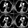
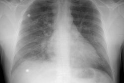
Clinically suspected pulmonary embolism: utility of spiral CT.
Kim KI, Muller NL, Mayo JR
PURPOSE: To prospectively determine the utility of contrast material-enhanced spiral computed tomography (CT) in the examination of patients clinically suspected of having pulmonary embolism (PE). MATERIALS AND METHODS: One hundred ten patients clinically suspected of having PE were examined with contrast-enhanced spinal CT and at least one other imaging modality: ventilation-perfusion scintigraphy, Doppler ultrasonography of deep leg veins, or pulmonary angiography. Chart review or telephone contact with the referring clinician was used to evaluate the contribution of spiral CT to the final clinical diagnosis. RESULTS: Spiral CT helped correctly identify 23 of 25 patients with PE (sensitivity, 92%). In 57 (67%) of the 85 patients without PE, spiral CT provided additional information that suggested or confirmed the alternate clinical diagnosis: pneumonia (n = 14), cardiovascular disease (n = 10), pulmonary fibrosis (n = 7), trauma (n = 6), malignancy (n = 5), pleural disease (n = 4), postoperative changes (n = 4), and other (n = 7). In the remaining 28 patients, spiral CT scans were normal (n = 12), failed to produce findings supportive of the final clinical diagnosis (n = 13), or were false-positive for PE (n = 3; specificity, 96%). CONCLUSION: Spiral CT has good sensitivity and specificity for the diagnosis of PE. In the majority of patients who do not have PE, it also provides important ancillary information for the final diagnosis.
PMID: 10207469, UI: 99223876


