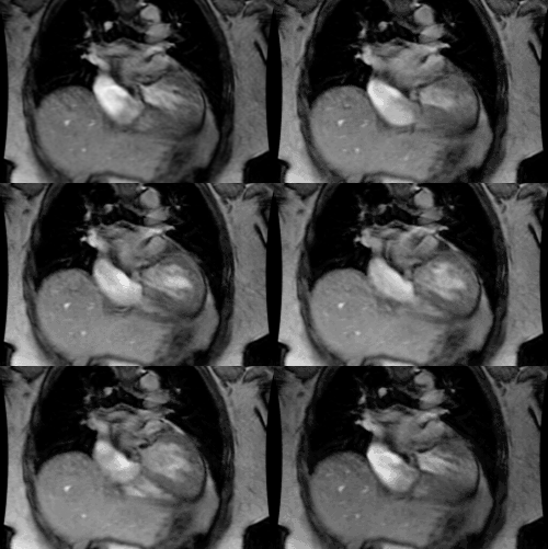
Situs and Heterotaxy Syndromes:
Situs refers to the positon of the visceral organs in the thoracic and abdominal cavities [5]. Situs colitus describes the normal anatomic position of the cardiac structures and thoracoabdominal organs [5]. Situs inversus refers to a left-right inversion of visceral and atrial positions in the thoracic and abdominal cavities (resulting in a mirror image of situs solitus) [5].Situs ambiguous is a term that characterizes positions of both atria and visceral organs in which lateralization across the left-right axis cannot be characterized as situs solitus or inversus, and is synonymous with left or right isomerism [5]. Isomerism describes the situation in which a persons morphologically right or left structures are found on both sides of the body [5]. Technically, any type of situs other than situs solitus can be described as heterotaxy, but the term heterotaxy is most commonly used in the context of situs ambiguous [5].Situs refers to the position of the atria (not the cardiac apex) and abdominal visera relative to the midline. Atrial situs is determined by identification of the morphologic right or left atrial structures. The morphologic right atrium receives the heptic veins, contains the crista terminalis, and the right atrial appendage (broad based triangular shaped and contains pectinate muscles that extend towards the atrioventricular valve [5]). The left atrial appendage is long and finger-like and contains pectinate muscles isolated to the body of the appendage [5]. Typically, the morphologic left atrium is on the same side as the aortic arch (but not always). When a coronary sinus is present, it always runs inferior-posterior to the morphologic left atrium and empties into the morphologic right atrium [5]. Cardiac situs solitus is present when a morphologic right atrium is located to the right of a morphologic left atrium [3]. Similarly, ventricular chambers must also be identified- the left ventricle has no infundibulum so there is fibrous continuity between the mitral and aortic valves. The left ventricle also has fine trabeculae and a smooth septal surface [5]. The right ventricle is usually trapezoidal in shape and the infundibulum separates the tricuspid from the pulmonary valve. The right ventricle also contains coarse trabeculae and a moderator band [5]. The orientation of the ventricles is commonly referred to as "ventricular looping" (in reference to the embryologic rotation of the cardiac tube during development) [5]. Rightward rotation is called D-loop and brings the future morphologic RV to its normal position rightward of the RV [5]. A left rotation (L-loop) brings the morphologic RV to the left of the LV [5].
Situs inversus represents a mirror-image configuration with a morphologic right atrium located to the left of a morphologic left atrium [3]. If the cardiac apex and gastric bubble are on opposite sides of the midline the patient has congenital heart disease until proven otherwise. Additionally, asplenia/ polysplenia must be ruled out.
Position of the cardiac apex:
1- Levocardia: Left sided apex (normal). Levocardia is not synonymous with situs solitus, although most patients with situs solitus have levocardia.
2- Dextrocardia: Cardiac apex is on the right. Most patients with situs inversus have dextrocardia.
3- Mesocardia: The bulk of the cardiac mass is in the center of
the
chest. There is no association with CHD and this condition is
usually of
no clinical significance.
Pulmonary situs: In situs abnormalities, the bronchopulmonary
anatomy is usually concordant with atrial situs [5]. The
morphologic right lung has an eparterial bronchus (the bronchus is
superior to the artery), while the left lung has a hyparterial
bronchus (the bronchus is below the pulmonary artery) [5]. The
morphologic right bronchus is typically more horizontal than the
left main bronchus and the right lung is trilobed, while the left
lung is bilobed [5].
Types of Situs:
1. Situs solitus with levoversion (Normal): Left sided left atrium; usually also have left cardiac apex and left aortic arch. Left hyparterial bronchus (bronchus runs beneath the left pulmonary artery) and right eparterial bronchus. Bilobed left lung; trilobed right lung. If the abdominal viscera are concordant with the thoracic structures (i.e.- left sided stomach and right sided liver) this is described as situs solitus totalis [3]. The incidence of CHD is about 0.8% [3].2. Situs inversus with mirror image dextrocardia: Situs inversus
with
dextrocardia represents the mirror image of situs solitus and is
the usual situation. There is left-right reversal of the
cardiac chambers coupled with a right apex and usually
right arch. There is ventricular inversion- the left ventricle
(smooth)
is located on the right, and atrial inversion- right atrium is
located
on the left and visa versa. The right ventricle (trabeculated)
remains
anterior and the left atrium remains posterior- ie: there is no
transposition
of the ventricular or atrial chambers. These patients have a
slightly increased
risk (3-5%) of congenital heart disease- most commonly corrected
transposition.
When there is reversal of the abdominal structures (right sided
stomach and left sided liver) this is referred to as situs
inversus totalis [3]. Twenty percent of patients with situs
inversus totalis have Kartagener's syndrome [3].Situs inversus
totalis is uncommonly associated with congenital heart defects
with an incidence of 2-5% [5].
3- Situs Indeterminatus (or situs ambiguous or heterotaxia or isomerism syndromes): See also Asplenia (Ivemark's syndrome) and Polysplenia
With true situs indeterminatus, there is abnormal arrangement of the organs and vessels. The stomach and liver are typically midline and congenital heart disease occurs in 50-100% of cases [2]. Major subcategories of situs ambiguous include- asplenia (right isomerism) and polysplenia (left isomerism)[2]. Minor forms of situs ambiguous include M-anisosplenia (third syndrome) and F-anisosplenia (fourth syndrome) [3]. These disorders are characterized by a bifurcated spleen (composed of one or more larger and one or more smaller splenunculi) and bronchial symmetry [3,4]. M-anisosplenia is more common in males and resembles a mild form of asplenia with bilateral eparterial bronchi and CHD (anomalous pulmonary venous return, pulmonary stenosis, common atrium, and bilateral SVC), but normal visceral situs [3,4]. F-anisosplenia is more common in females and resembles a severe form of polysplenia with bilateral hyparterial bronchi (bilateral left lung) and abnormal visceral situs (especial intestinal malrotation) [3,4]. Associated cardiac anaomalies include double-outlet right ventricle, bilteral SVC, and azygous continuation of the IVC [3].
A. Dextroversion: Situs solitus with dextrocardia. Gastric bubble and spleen are on the left, the liver is on the right, and the cardiac apex is on the right. Generally, the aortic arch is left sided. There is NO chamber inversion- right ventricle and atrium remain on right side, and left ventricle and atrium remain on the left side; however, the normal anterior to posterior chamber relationships are lost (right ventricle becomes the posterior ventricular chamber)- ie: there is transposition of the cardiac chambers. Congenital heart disease is found in 95% of cases. Congenitally corrected transposition occurs with increased frequency in these patients.
B. Levoversion: Situs inversus with levocardia. Characterized by: Gastric bubble and spleen are on the right, and the liver on the left (situs inversus). There is typically a right arch. The majority of the cardiac mass is in the left chest, but the apex is due to the right ventricle. Chamber inversion (reversed left and right relationships): the right ventricle (atrium) is on the left and forms the cardiac apex (which will be left sided). Left ventricle (atrium) is on the right. Chamber transposition (reversed anteroposterior relationships): The right ventricle (atrium) will be the posterior ventricular (atrial) chamber. Levoversion is ALWAYS associated with congenital heart disease. Also must exclude asplenia/ polysplenia syndromes.
REFERENCES:
(1) Radiographics 1999; Applegate KE, et al. Situs revisited:
Imaging
of the heterotaxy syndrome. 19: 837-852
(2) Radiographics 2002; Fulcher AS, Turner MA. Abdominal manifestations of situs anomalies in adults. 22: 1439-1456
(3) AJR 2009; Ghosh S, et al. Anomalies of visceroatrial situs. 193: 1107-1117
(4) AJR 1975; Landing BH. Syndromes of congenital heart disease
with tracheobronchial anomalies. 123: 679-
(5) J Cardiovasc Comput Tomogr 2013; Wolla CD, et al.
Cardiovascular manifestations of heterotaxy and related situs
abnormalities assessed with CT angiography. 7: 408-416


