Takayasu's Arteritis:
Clinical:
Takaysu's arteritis is a large vessel vasculitis that produces destruction of the arterial media and affects elastic arteries that possess a vasa vasorum [8]. It typically affects the aorta and it's proximal branches [8]. The pulmonary arteries are involved in 15% of patients [11]. Muscular peripheral arteries are not involved. The disorder is characterized by a granulomaotous inflammation of the arterial wall (inflammation of the vasa vasorum of the adventitia and lymphocytic infiltration of the media [12]) with marked intimal proliferation and fibrosis of the media and adventitia [8]. This eventually leads to stenosis and vessel occlusion, but occasionally there can be poststenotic dilatations and aneurysm formation (when the inflammation destroys the media) [6,8]. The lesions tend to be segmental with a patchy distribution [8].
Takayasu's affects females more than males (10:1)- especially
young Asian women, generally in the 2nd or 3rd decade of life [9].
The onset of the disorder is insidious and it can be divided into
an early "pre-pulseless" phase and a late occlusive phase [6].
Early patients may experience fever, night sweats, weakness,
arthralgia, myalgia, and a skin rash. Laboratory abnormalities
include an elevated ESR, anemia, and a positive C-reactive protein
test. Unfortunately, ESR can be normal in up to one-third of
patients with active disease and remain persistent elevated in up
to 56% of patients in remission [5]. Later, patients present with
occlusive symptoms such as bruits, diminished or absent pulses,
renal vascular hypertension, and cerebrovascular or visual
complaints. The time interval between early and late phases may
vary from one to eight years, however, various phases may coexist
at the same time [8]. Early diagnosis is important as the early
initiation of steroid therapy can result in dramatic improvement
in the clinical and radiologic abnormalities. Steroids induce a
remission of disease in 50% of patients, with the other 50%
responding well to methotrexate [12].
The disorder most commonly involves the thoracic aorta, its branches, and the pulmonary arteries (pulmonary arteries are involved in 50-80% of cases and this is often a late manifestation of the disease) [7]. The most characteristic findings are stenosis or occlusion, mainly of the segmental and subsegmental arteries and less commonly of the lobar or main pulmonary arteries [7]. Pulmonary involvement is infrequently associated with symptoms or functional compromise. [1] Up to 60% of cases of Takayasu arteritis can demonstrate coronary artery involvement (which is higher than that seen in giant cell arteritis) and 5-20% of patients will have clinical symptoms such as myocardial ischemia [13].
Takayasu's is classified according to the site of involvement [10]:
Type I- only supraaortic artery involvement
Type IIa - only ascending aorta or aortic arch
Type IIb- descending aorta, with or without the more proximal portions of the aorta and aortic arch branches
Type III- aortic arch and it's branches with simultaneous involvement of the descending thoracic and abdominal portion or renal arteries
Type IV- only abdominal aortic or renal artery involvement
Type V- generalized involvement as a combination of the other types
Coronary and pulmonary artery involvement are indicated as C(+) or P(+)
Complications include development of aortic aneurysm which can be seen in up to 45% of affected patients [4].
X-ray:
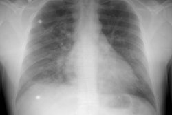
Plain film: Early, the shadow of the descending thoracic aorta can become "wavy" or "scalloped". Later findings include linear calcification in the aortic arch and descending aorta (the ascending aorta is spared) and decreased pulmonary vascular markings.
Ultrasound: The typical sonographic finding is a long, smooth, homogenenous circumferential thickening of the arterial wall0 the so-called "halo sign" [10].
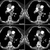
Computed Tomography: On CT, the wall of the thoracic aorta becomes thickened (more than 1 mm). On CECT, the wall demonstrates a double ring pattern with a low density inside ring (due to mucoid swelling of the intima) and an enhancing outer ring (felt to represent active medial and adventitial inflammation). Pulmonary artery involvement appears as dilatation, diffuse wall thickening, or a high-attenuation within the wall on non-contrast exam [3]. Increased attenuation can be seen in the mediastinal fat surrounding the aorta and pulmonary arteries due to active inflammation [11]. Parenchymal findings include a mosaic pattern with areas of decreased attenuation containing a reduced number and caliber of vessels which correspond to areas of hypoperfusion angiographically [2]. Other findings include areas of mild to moderate pleural thickening and subpleural reticulolinear and patchy changes [2]. In the chronic phase- mural calcium deposition and lumenal stenosis or occlusion occur [8].
MRI: Delayed vessel wall enhancement is a finding that can be identified in patients with Takayasu's [5].
FDG PET: Abnormal tracer accumulation can be seen in the walls of the involved vessels [10].
Angiography: Angiography can be used to demonstrate the lumenal changes of the disorder such as stenosis, occlusion, or aneurysmal dilatation of the involved vessels. Stenosis is the most common abnormality- frequently involving the descending thoracic and abdominal aorta, the subclavian, common carotid, and renal arteries. Narrowing can be short and segmental or long and diffuse. Occlusions (characteristically "flame-shaped") can also occur and dilated collateral vessels will usually be detected. Aneurysmal dilatation (either fusiform [most commonly] or saccular) may also be seen and is more common in the ascending aorta, and the right brachiocephalic and right subclavian arteries. Segmental septa of the carotid artery are characteristic of the disorder and represent multisegmental dilatations separated by regions of normal-appearing non-dilated vessel.
Pulmonary involvement may result in the development of bronchial-pulmonary artery shunts.
REFERENCES:
(1) Radiographics 1997; 17: 579-594
(2) J Comput Assist Tomogr 1996; Takahashi K, et al. CT findings of pulmonary parenchyma in Takayasu arteritis. 20: 742-748
(3) AJR 1999; Jean-Francois P, et al. Electron beam CT features of pulmonary artery in Takayasu's arteritis. 173: 89-93
(4) AJR 2000; Sueyoshi E, et al. Aortic aneurysms in patients with Takayasu's arteritis: CT evaluation. 175: 1727-1733
(5) AJR 2005; Desai MY, et al. Delayed contrast-enhanced MRI of the aortic wall in Takayasu's arteritis: initial experience. 184: 1427-1431
(6) AJR 2005; Gotway MB, et al. Imaging findings in Takayasu's arteritis. 184: 1945-1950
(7) Radiographics 2006; Castaner E, et al. Congenital and acquired pulmonary artery anomalies in the adult: radiologic overview. 26: 349-371
(8) Radiographics 2010; Castaner E, et al. When to suspect pulmonary vasculitis: radiologic and clinical clues. 30: 33-53
(9) Radiographics 2010; Kimura-Hayama ET, et al. Uncommon congenital and acquired aortic diseases: role of multidetector CT angiography. 30: 79-98
(10) AJR 2010; Spira D, et al. Imaging of primary and secondary inflammatory diseases involving medium-sized vessels and their potential mimics: a multitechnique approach. 194: 848-856
(11) Radiology 2010; Chung MO, et al. Imaging of pulmonary
vasculitis. 255: 322-341
(12) AJR 2013; Nemec SF, et al. Noninfectious inflammatory lung
disease: imaging considerations and clues to differential
diagnosis. 201: 278-294
(13) Radiographics 2018; Broncano J, et al. CT and MR imaging of
cardiothoracic vasculitis. 38: 997-1021


