AI based lesion-detection software used in daily practice can help identify clinically significant incidental lung nodules on chest x-rays, according to a study published November 13 in Scientific Reports.
A group at Yonsei University College of Medicine in Gyeonggi-do, South Korea, evaluated how often clinically significant lung nodules were detected unexpectedly on chest x-rays by AI (Insight CXR, v3, Lunit) and whether coexisting findings can aid in differential diagnoses.
“Our results showed that lung nodules were detected unexpectedly by AI in approximately 1% of initial [chest x-rays], and approximately 70% of these cases were true positive nodules, while 20.5% needed clinical management,” wrote lead author Shin Hye Hwang, MD, and colleagues.
Insight CXR has been used as an aid to help interpret all posterior-anterior and anterior-posterior chest x-rays at the authors’ hospital since March 2021, with the group continually evaluating its impact in clinical practice. The software is designed to detect eight types of lesions, including nodules, pneumothorax, consolidation, atelectasis, fibrosis, cardiomegaly, pleural effusion, and pneumoperitoneum.
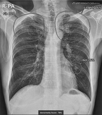
When patients undergo a chest x-ray, the software automatically analyzes the image and attaches a secondary file to the original image in the hospital’s PACS. Clinicians then may refer to the AI results, which are displayed with a contour map, abbreviations, and an abnormality score.
 A true positive case in a 72-year-old male with a granuloma on chest x-ray after visiting the neurology outpatient clinic for memory disturbance. A small lung nodule was detected in the left-middle to lower lung field by AI software, with an abnormality score of 26%. Co-existing fibrosis was suspected in the apex of bilateral upper lungs, with an abnormality score of 76%. Image courtesy of Scientific Reports.
“This allows for real-time utilization of AI-generated results alongside patient imaging in our hospital,” the authors noted.
In this study, the researchers evaluated imaging results from 14,563 patients who underwent initial chest x-rays in outpatient clinics. Three radiologists categorized nodules into malignancy (group A), active inflammation or infection that needs treatment (group B), (group C), and others (group D). Lesions were considered present when the software’s abnormality score exceeded 15%.
According to the findings, the AI software detected unexpected lung nodules in 152 patients (1%). The study authors excluded 72 patients due to inconclusive results because they had no follow-up images. In addition, seven patients were excluded because they received no final clinical diagnosis.
Of the 73 patients included in the final analysis, the false positive rate was 30.1%. The proportion with malignancy was 11%, active inflammation was 6.9%, postinflammatory sequelae was 49.3%, and others was 2.7%. This indicates that approximately 20.6% of incidental lung nodules of group A, B, and D required further evaluation or treatment, the group wrote.
Ultimately, the authors noted that there remains a lack of information on detection and management of lung nodules depending on use of AI-based software. In their hospital, clinicians can assess the AI results whenever they want, and thus the researchers were not able to verify whether the decision-making process involved included reference to the AI results.
However, they wrote that they plan to address these issues in a subsequent study.
“Further research is needed in collaboration with hospitals that have not yet introduced AI-based detection software,” the group concluded.
The full article is available here.


