An investigational new PET radiotracer guided more curative surgeries than standard imaging methods for patients with primary hyperparathyroidism, according to research published online July 21 in Surgery.
The study authors analyzed the clinical impact of PET imaging with a radiotracer called F-18 fluorocholine (FCH) for guiding parathyroid surgery in patients with cancer and compared the results with standard imaging with ultrasound or sestamibi scintigraphy. They found that F-18 FCH-PET was more accurate at identifying tumors and subsequently led to more surgical "cures."
"Long-term results in the first cohort in the United States to use 18F-fluorocholine positron emission tomography for parathyroid localization confirm its utility in a challenging cohort, with better sensitivity than ultrasound or sestamibi," wrote lead author Dr. Claire Graves, an endocrine surgery fellow at University of California San Francisco, and colleagues.
Primary hyperparathyroidism is one of the most common hormonal disorders, with women affected three to four times more often than men. Approximately 100,000 people develop the condition in the U.S. each year, and surgical resection of abnormal glands is the mainstay for curative treatment.
Confident and accurate preoperative localization by noninvasive imaging studies is an important and challenging issue for successful parathyroidectomy. Although new imaging modalities have been introduced during the past decade, direct comparative studies on advanced imaging techniques are limited, according to the authors.
In this study, a first analysis on the effect of FCH-PET on clinical decision-making in the U.S., the researchers aimed to provide a clinically relevant assessment of FCH-PET parathyroid localization.
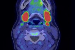
They performed imaging on 101 candidates for parathyroid surgery with F-18 FCH radiotracer using a 3.0 Tesla time-of-flight PET/MRI system (GE Healthcare). In patients who underwent subsequent surgery, cure was based on lab values at least six months after surgery. Location-based sensitivity and specificity of F-18 FCH-PET were also assessed using three anatomic locations: the left neck, right neck, and mediastinum.
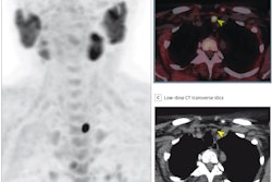
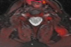
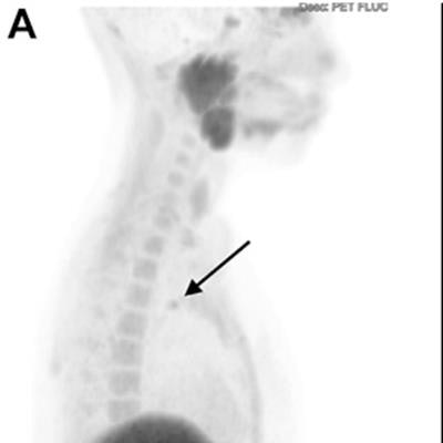
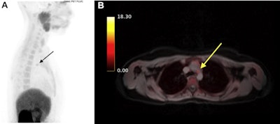 FCH-PET image of an ectopic mediastinal parathyroid adenoma (arrows) on (A) sagittal and (B) axial view fused with simultaneous MRI. This patient had previously undergone a bilateral neck exploration, which had failed to localize a left lower gland. Ultrasound, sestamibi, and 4D CT were all negative. After FCH-PET localization, a robot-assisted left thoracoscopic approach was employed to resect the gland from the aortopulmonary window. Follow-up blood work 8 months after surgery confirmed normalization of calcium. Image courtesy of Surgery.
Graves and colleagues found that F-18 FCH-PET localized at least one cancer lesion in 93% of patients overall and in 91% of patients with previously negative imaging, leading to a change in preoperative strategy in 60% of patients. Of 76 patients who underwent parathyroidectomy, 58 (77%) had laboratory data at least six months afterward, with 55 out of 58 of these patients (95%) demonstrating cure.
In addition, F-18 FCH-PET successfully guided more curative surgery and had higher sensitivity than ultrasound and sestamibi imaging.
| Performance of F-18 FCH-PET for guiding parathyroid surgery | |||
| Sestamibi scintigraphy | Ultrasound | FCH-PET | |
| Curative surgery | 13/55 (24%) | 20/57 (35%) | 48/58 (83%) |
| Sensitivity | 27.5% | 37.1% | 88.9% |
"PET imaging demonstrated markedly superior sensitivity compared with both [ultrasound] and sestamibi," the researchers wrote.
FCH is not yet approved by the U.S. Food and Drug Administration or widely commercially available, the authors noted. A phase II clinical trial evaluating F-18 FCH-PET for localizing parathyroid adenomas in patients with hyperparathyroidism prior to surgery is ongoing, with an estimated completion date in December 2021.
Thus, the costs of these studies are not yet known. They will likely be expensive, however, according to the authors.
"Future analysis of the added value and cost-effectiveness of FCH PET will be crucial to define the role of this imaging in patient care," Graves et al concluded.


