F-18 fluorothymidine (FLT) PET can identify early acute gastrointestinal graft versus host disease (GVHD) after patients undergo bone marrow transplants, according to a study published December 13 in Radiology: Imaging Cancer.
The finding is from a clinical trial in patients with blood cancers and suggests the method can locate specific areas of the disease that are difficult to biopsy, noted lead authors Jennifer Holter-Chakrabarty, MD, and Lacey McNally, PhD, of the University of Oklahoma Health Sciences Center in Oklahoma City.
“Severe acute GVHD continues to confer increased transplant-related mortality, and there are currently no biomarkers to identify those at risk to develop this disease,” the researchers wrote.
Blood cancers start in the cells of the immune system or in blood-forming tissue, such as the bone marrow. Hematopoietic stem cell transplantation (HSCT) is an effective treatment, but there is a risk that the new donor’s immune system will not recognize the host tissues and subsequently attack them. This is known as GVHD and can be life-threatening, the authors explained.
Diagnosis of GVHD is usually based on elevated levels of blood cells secreted by inflammatory lymphocytes, but requires invasive methods such as biopsy to confirm, the authors noted. Moreover, gastrointestinal areas are difficult to biopsy, the researchers noted. Conversely, F-18 FLT-PET imaging can capture the proliferation of lymphocytes, including those that may drive GI-GVHD, and in this study, the group hypothesized that the approach could identify the disease based on uptake of F-18 FLT radiotracer in the GI system.
To test the hypothesis, the researchers analyzed F-18 FLT-PET standardized uptake values (SUVs) in GI regions in 20 patients (median age, 34 years old, 11 women, nine men) who underwent transplantation. Seven of the patients developed biopsy-confirmed GI-GVHD by day 100 after the procedure. F-18 FLT-PET imaging was performed on day 28.
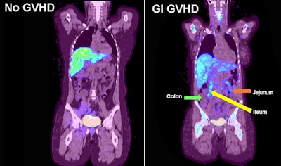
 F-18 FLT-PET imaging of gastrointestinal GVHD. Examples of F-18 FLT uptake in a participant with no GVHD (left) and a participant with acute GVHD and uptake in the jejunum and ileum (right). Arrows denote increased uptake in the GI tract. Image available for republishing under Creative Commons license (CC BY 4.0 DEED, Attribution 4.0 International) and courtesy of RSNA.
In the seven patients, the researchers specifically analyzed regions of interest in the colon, jejunum, and ileum. F-18 FLT-PET SUV in the jejunum in participants with acute GVHD was increased (median maximum SUV, 4.19) compared with participants without GVHD (SUVmax, 2.43), according to the findings.
In addition, for each increase of one point in the F-18 FLT-PET SUVmax in the jejunum, the odds of being diagnosed with GI-GVHD were 12.58 times greater, the researchers noted. The SUV uptake (mean, maximum, and total) in the colon and ileum of the acute GVHD group was higher than the non-GVHD group but was not statistically significant.
“This study suggests that the jejunum may be an underappreciated site of GI-GVHD and that increased uptake at this site may identify participants before the onset of clinical symptoms,” the group wrote.
Ultimately, the study supports future larger imaging studies using F-18 FLT-PET and serum biomarkers that may predict and diagnose GI-GVHD early after HSCT, the group concluded.
The full study can be found here.


