F-18 FDG-PET/CT is a valuable approach for diagnosing unknown infections in intensive care unit (ICU) patients, according to research published February 20 in Annals of Intensive Care.
The finding is from a study in two medical centers in the Netherlands in which patients with persistent illness underwent at least seven other types of diagnostic procedures prior to the PET/CT scans, including x-rays and CT scans, noted lead author Bram van Leer, MD, of the University of Groningen, and colleagues.
“Remarkably, F-18 FDG-PET/CT identified an infectious or inflammatory focus in 92% of patients, with 72% of those reported to be new,” the group wrote.
Some ICU patients remain critically ill due to hospital-acquired infections despite a reversal of their original diagnosis, and outcomes for these patients are poor, with an estimated death rate of 25% due to organ failure, the authors explained. While F-18 FDG-PET/CT is routinely used outside of critical care settings in patients with bacteremia, fever of unknown origin, vascular graft infections, or joint infections, for instance, its use in ICUs has been described in only a few small single-center observational studies, they noted.
To explore the additional value of the technique, the group studied clinical reports on 47 patients admitted to ICUs between 2017 and 2024 who underwent F-18 FDG-PET/CT scans ten days or more after admission. They determined whether diagnoses were new, which diagnostics were performed before the PET/CT scans, and whether PET/CT resulted in treatment changes.
Among the patients, respiratory support was the most common primary reason for prolonged length of ICU stay (n = 26) and during the PET/CT scans, most patients were on mechanical ventilation (n = 39) and/or circulatory support (n = 35). The median interval between admission and PET/CT was 21 days, the authors noted.
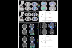
 There are several conceivable advantages of F-18 FDG PET/CT over conventional imaging. Image A and B showing an example of the distinctive capability of the F-18 FDG PET/CT. A shows a fusion image of F-18 FDG PET/CT and low-dose CT and B an high resolution chest CT of a patient with an Aspergillus infection. The suspected aspergilloma is concealed on the high resolution CT due to the lung fluids and consolidation. Image C and D (fusion image of F-18 FDG PET/CT and low-dose CT and low-dose CT only, respectively) are examples of the ability to image inflammation while no concurrent abnormalities are visualized, in this case prostatitis (red arrows). Image E, a maximized intensity projection of F-18 FDG PET, shows a patient with lung infections and multiple muscle abscesses after MRSA sepsis. This is an illustrative example of a relevant dissemination investigation. Image and caption available for republishing under Creative Commons license (CC BY 4.0 DEED, Attribution 4.0 International) and courtesy of Annals of Intensive Care.
According to the results, PET/CT helped detect a potential infectious or inflammatory focus in 43 patients (91%), of which 34 (72%) were previously unknown. The scans were performed late in the diagnostic workup, with a median of seven diagnostic procedures performed prior to PET/CT, and in 26 (55%) patients, therapy changes were reported within 48 hours after PET/CT, the authors reported.
“In more than half of the included patients, PET/CT contributed to a change in therapy, highlighting its potential role in guiding clinical decisions within the ICU,” the group wrote.
Ultimately, PET/CT scans are considered complex procedures for critically ill patients, the authors noted. However, with new large field-of-view PET/CT scanners available, whole-body scans can be acquired within a couple of minutes, they wrote. Well-structured preparation protocols and better awareness among ICU personnel could thus increase adoption of F-18 FDG PET/CT imaging in ICU patients, the authors suggested.
To the best of their knowledge, the study is the first multi-center and largest to report on the potential value of F-18 FDG PET/CT imaging in critically ill patients, the group added.
“Future studies should investigate if biomarkers can predict [patients] with high diagnostic yield from PET/CT. In addition, direct comparisons between F-18 FDG-PET/CT and contrast-enhanced CT should be investigated,” the group concluded.
The full study is available here.