A new PET radiotracer rapidly detected and localized life-threatening mold-related invasive fungal infections, according to a presentation June 23 at the 2025 Society of Nuclear Medicine and Molecular Imaging (SNMMI) annual meeting.
Researchers looking for ways to leverage the widely used F-18 fluorodeoxyglucose (F-18 FDG) radiotracer say that F-18 fluorodeoxysorbitol (F-18 FDS) can be easily produced from F-18 FDG and synthesized on demand. Most notably, F-18-FDS shows promise as a noninvasive diagnostic tool for addressing life-threatening mold-related invasive fungal diseases (IFDs), according to postdoctoral research fellow Carlos Ruiz-Gonzalez, MD, and colleagues from Johns Hopkins University Medical School in Baltimore, MD..
The findings are important because invasive mold infections have become increasingly common in people who have undergone cancer and immunosuppressive treatments.
“Currently, it’s very difficult to detect invasive mold infections,” Ruiz-Gonzalez said in SNMMI statement. “Definitive diagnosis often depends on invasive procedures or on biomarkers that lack sensitivity for many mold species."
To explore the potential value of F18-FDS, the team first tested in vitro uptake of the radiotracer to determine its ability to detect 30 different strains of disease-causing molds collected from infected patients. Next, they performed PET/CT imaging on immunosuppressed mice, four human patients with confirmed invasive mold infections, and five control patients with inflammatory diseases or cancer, but no infections.
In the prospectively enrolled patients, F-18 FDS PET whole-body imaging detected and localized invasive pulmonary and cerebral infections in all patients with mold infections [Aspergillus, non-Aspergillus (galactomannan-negative) molds, or azole-resistant strain] with a target-to-nontarget ratio > 3, and differentiated them from sterile inflammation and cancer (p < 0.01), the team reported.
In the mouse studies, F-18 FDS PET localized invasive lung and brain Aspergillosis, as well as sinus infections related to Aspergillus, Rhizopus, and Mucor, according to the results. The technique also identified a cerebral lesion previously missed on a brain MRI of a patient with fungal Cladophialophora bantiana pulmonary infection.
In addition, F-18-FDS PET/MRI allowed for the precise localization of fungal infection sites within the cerebral parenchyma and enabled correlation with lesions found on histology, the team also reported.
The group noted that while Enterobacterales also metabolize sorbitol, tissue histology from eight patients with invasive mold infections demonstrated that molds constitute a higher proportion (11.7%) of infected lesions than bacterial infections (0.24%), leading to a substantially higher F-18 FDS PET signal from mold versus bacterial infections in vivo.
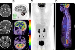
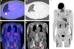
 Representative F-18 FDS PET/CT images of patients with (a) pulmonary aspergillosis (n = 3), (b) control patient with breast cancer (n = 1). In this patient, F-18 FDG PET shows the cancer lesion (arrow) which is not visualized on the F-18 FDS PET. (c) Control patients with interstitial lung disease (n = 4). (d) Quantification of F-18 FDS PET as target-to-nontarget (TNT) ratio; n = 3 volume of interest per patient. Representative F-18 FDS PET/CT images of a patient with (e) invasive cerebral aspergillosis and (g) uninfected control. (f) Transverse sections showing MRI and PET/MRI from a patient with invasive cerebral aspergillosis. (h) F-18 FDS PET quantification TNT ratio in brain derived from two patients with invasive cerebral mold infections, and five control patients without cerebral pathology with n = 2 VOIs per patient (patient 4 with mold infection has 1 VOI only). Lesions are highlighted with arrows.SNMMI
Formulating F-18 FDS using a one-step kit method may allow for on-demand synthesis and global availability of the novel radiotracer, ultimately leading to prompt identification of IFDs, according to the group.